Rebuilding Alan's Thumb

By Whitney Lowe
[Clinical Apps]

Clinical massage practitioners are most successful when they don’t jump to immediate conclusions, but take the time to fully investigate the nature of a client’s problem. In this installment of Clinical Apps, we explore a case in which the client is convinced he has one condition, but in fact it is something else. It is through your developed critical thinking and problem-solving skills that you will find effective solutions to your clients’ problems.

Background and Key Considerations
Alan recently took a carpentry job in which he uses a lot of power tools. He is suffering hand pain that interferes with his work. He is seeking health care because he is concerned that his symptoms may be a result of carpal tunnel syndrome.  
Alan’s primary complaint is sharp pain around the wrist and base of the thumb. In certain positions, the pain is sharp enough to make him feel like he is going to drop the items he is holding. He reports that this sensation is not so much an actual loss of overall grip strength, but a sudden, sharp pain around the base of the thumb, resulting in an abrupt loss of hand and grip strength.
In assessment, the essential rule of real estate applies: location, location, location. Of course, the site of pain does not always correspond with the location of the actual tissue dysfunction, as in trigger-point referral or radiating nerve pain, but pain location does provide a good clue and starting point for evaluation.
Carpal tunnel syndrome is common among carpenters due to the job’s constant gripping actions and vibrations from power tools. These are key factors causing median nerve compression under the flexor retinaculum. However, just because Alan has hand pain and carpal tunnel syndrome is common in his profession doesn’t mean we can jump to the conclusion that it is the source of the present complaint. Other key structures in the area need to be investigated.
Multijoint tendons that control the thumb and fingers have several unique factors that can lead to pain complaints in this area. The thumb and finger tendons that cross the wrist all pass under fibrous restraints called retinacula (singular: retinaculum). Each retinaculum plays a key biomechanical role in force transmission for the tendons. Looking at the retinacula, it would appear that their primary function is to hold the tendons in place around the joint. While they do provide a restraining force for the tendons, they actually perform a more critical biomechanical role.
There are unique biomechanical challenges for tendons that cross multiple joints, like those of the wrist and fingers. For example, in the distal extremities, tendons often exert their pulling force across a 90-degree angle. Generating force across a sharp angle like this requires a mechanical arrangement like a pulley and rope arrangement. The retinaculum acts like a pulley so the tendons can continue to generate a strong force across this sharp angle.
Unfortunately there is a serious downside to the retinacula’s role as a pulley, which should be apparent. If the tendon is pulling strongly against the pulley (retinaculum), there is a great deal of pressure and friction between the tendon and retinaculum. Increased friction on the tendon can lead to pain and tendon degeneration.
The body compensates for the increased friction by enclosing the tendon in a synovial sheath. The sheath is in contact with the retinaculum, but the tendon slides back and forth inside the sheath and thereby reduces friction between the tendon and the binding retinaculum.
The tendon sheath greatly reduces stress on the tendons; however, they are not immune from pathology due to overuse. Chronic overuse can cause an inflammatory reaction between the tendon and surrounding synovial sheath, causing fibrous adhesions to develop there as a result of the irritation. Having inflammation and fibrous adhesions between the tendon and surrounding sheath is called tenosynovitis.
There is a significant distinction between tenosynovitis and tendinosis (often called tendinitis), which is the most common tendon pathology. Tendinosis is not an inflammatory condition—it results from collagen degeneration within the tendon. Tenosynovitis, on the other hand, does have inflammatory activity in the tissues, although it is frequently not apparent during physical examination.
Alan reported pain in the wrist, near the base of the thumb. This is a location where two thumb tendons are highly susceptible to tenosynovitis; a condition called de Quervain’s tenosynovitis. The profile created by the two tendons when their muscles are contracted is sometimes referred to as the anatomical snuff box. These two thumb tendons, the abductor pollicis longus and extensor pollicis brevis, course beneath a binding retinaculum (Image 3). The extensor pollicis longus tendon is also in this region, but does not cause as many problems. Numerous occupational activities put stress on the thumb tendons underneath the retinaculum here. In fact, tenosynovitis in these thumb tendons is a common injury that plagues massage therapists, especially if they perform a large amount of thumb-pressure work, which many do.
The exact cause of tenosynovitis is not always clear, but there does seem to be a strong correlation between overuse and the condition’s development. Repeated friction is clearly a causative factor. A recent study unsurprisingly found a strong correlation between the onset of de Quervain’s tenosynovitis and high-volume texting with the thumbs on a smartphone.1  
Some anatomical anomalies occur in this region, including a fibrous septum between the abductor pollicis longus and extensor pollicis brevis muscles. This fibrous division between the muscles may narrow the channel through which the tendons pass and increase friction leading to tenosynovitis. This septum is not palpable through physical examination and is only evident with high-tech diagnostic studies.
Many references to de Quervain’s tenosynovitis refer to it as stenosing tenosynovitis. Stenosis means narrowing, and stenosing tenosynovitis refers to a narrowing of the channel through which the tendon passes. Sometimes fibrous nodules develop along a tendon right where the tendon passes beneath a retinaculum. The fibrous nodule causes the tendon to get stuck as it attempts to slide under the retinaculum. This pathology occurs most often in the flexor tendons of the fingers and is commonly referred to as trigger finger.

Assessment and Evaluation
Carpal tunnel syndrome and de Quervain’s tenosynovitis can produce pain in a similar location. Yet, there are some key differences that help discriminate these conditions. The pain in carpal tunnel syndrome is more common in the palm of the hand, not the base of the thumb as Alan reports. Pain at the base of the thumb is more consistent with tenosynovitis.
Palpating the tendons in the anatomical snuff box region reproduces Alan’s primary complaint. Pain is significantly increased if the tendons are palpated while their associated muscles are being contracted. Many tendon complaints are more pronounced when there is a contraction force on the muscle-tendon unit. If median nerve compression was the culprit, pain reproduction in this region would be unlikely because pressure is not applied over the nerve. It is also unlikely that pain in that region would increase with the muscle contraction if it were median nerve compression.
Alan’s pain is also reproduced with resisted extension and resisted abduction of the thumb. These are the two key motions of the involved tendons. In addition to investigating motions of the thumb, other wrist motions are assessed. There was no pain associated with active or passive flexion of the wrist. In carpal tunnel syndrome, these motions generally reproduce symptoms because they compress the median nerve in the carpal tunnel.
Another key factor that points to the likelihood of tenosynovitis is pain reproduced with a special orthopedic test called the Finklestein test. It is a simple procedure where the client pulls his thumb across his palm and then wraps his fingers over the thumb. With the thumb and fingers in that position, the wrist and hand are moved in ulnar deviation. If this position reproduces the primary pain complaint, it is likely there is some involvement with the key thumb tendons and/or their synovial sheaths. This testing procedure does not stress the median nerve at all, so we can rule out carpal tunnel involvement in favor of a tendon overuse disorder (unless other symptoms indicate both conditions).

Treatment Considerations
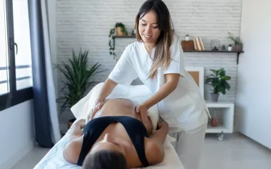
The first step in our clinical management of any soft-tissue disorder is to normalize the soft-tissue dysfunction; in this case inflammatory irritation and fibrous adhesions between the tendon and surrounding synovial sheath. In any chronic overuse tendon disorder, it is crucial to reduce the cumulative tensile load on the tendon by working on the affected muscles to decrease their tightness as much as possible. The muscle bellies of the associated muscles extend quite a distance into the forearm, and should be worked throughout their entire length (Image 5).
After initial tissue warming and superficial strokes are applied, deep specific stripping applications are applied to the bellies of the abductor pollicis longus and extensor pollicis brevis (Image 6). Pressure can increase in successive strokes as the client is able to tolerate it. The extensor pollicis longus should be treated with the same stripping techniques at this time as well.
Following the initial stripping techniques, perform those same stripping techniques simultaneously with movement that lengthens the muscle. Use one hand to perform the stripping technique, and the other hand to pull the client’s hand into ulnar deviation, just like in the Finklestein test. Stretching the muscle-tendon unit in this direction as the stripping technique is applied is highly effective in achieving the best muscle relaxation for the thumb muscles.
Recent studies have highlighted the benefits of conservative soft-tissue manipulation techniques in treating tenosynovitis.2 Some of these techniques are performed with the hands, while others such as gua sha and Graston technique are performed with a specific tool. There is a fair amount of controversy about some of the instrument-assisted soft-tissue treatments because they can appear to cause tissue damage and bruising. However, evidence suggests they are effective, so further research is warranted. Only those skilled in these techniques should use them.
Treating the key muscle bellies will allow them to relax, and will reduce the load on the tendon. Attention can now focus on addressing the specific tendon dysfunction. Deep-friction massage to the affected tendon is the treatment of choice. The primary purpose of friction to treat tenosynovitis is to address the fibrous adhesions and encourage tendon healing.
Friction treatment is applied to the damaged tendon to stimulate fibroblast proliferation, which will encourage tissue repair. Studies have shown that the key to effective fibroblast stimulation is pressure and movement applied to the tendon. The friction can be applied in different directions (longitudinally or transverse) and still produce beneficial effects.3 However, sometimes transverse friction is more effective in breaking up fibrous adhesions that develop between the tendon and its surrounding synovial sheath.
Putting the tendon on a stretch when friction is applied also increases the effectiveness of the technique. With the tendon pulled taut, the pressure is more effectively delivered to the tendon and it can help break up any fibrous adhesions.
Tenosynovitis treatments are most effective when they are performed frequently. The more often the pressure and movement are applied to the tendon, the more effective the treatment. When there is tenosynovitis in the thumb tendons, as in Alan’s case, massage treatments that are applied to the entire upper extremity are also valuable. It will be important for Alan to continue his treatments for long-term resolution. However, Alan can also be taught to perform these friction treatments on himself, and the more frequently he does the techniques, the faster he can restore optimum function.

Conclusion
Clients may often come in with preconceived fears or concerns that they have a particular pathology, especially if it is something extremely common like carpal tunnel syndrome. However, as in Alan’s case, sometimes our analytical skills help clarify the real nature of the problem through evidence-based rationale. When you do this, you increase the confidence your client places in you, while greatly improving your treatment success at the same time.  

Notes
1.    Maryam Ali et al., “Frequency of De Quervain’s Tenosynovitis and Its Association with SMS Texting,” Muscles, Ligaments and Tendons Journal 4, no. 1 (2014): 74–8.
2.    John A. Papa, “Conservative Management of De Quervain’s Stenosing Tenosynovitis: A Case Report,” Journal of the Canadian Chiropractic Association 56, no. 2 (2012): 112–20; Emily R. Howell, “Conservative Care of De Quervain’s Tenosynovitis/Tendinopathy in a Warehouse Worker and Recreational Cyclist: A Case Report,” Journal of the Canadian Chiropractic Association 56, no. 2 (2012): 121–7;  Warren I. Hammer, “The Effect of Mechanical Load on Degenerated Soft Tissue,” Journal of Bodywork and Movement Therapies 12, no. 3 (2008): 246–56.
3.    G. M. Gehlsen, L. R. Ganion, and R. Helfst, “Fibroblast Responses to Variation in Soft Tissue Mobilization Pressure,” Medicine & Science in Sport & Exercise 31, no. 4 (1999): 531–35.

Whitney Lowe is the author of Orthopedic Assessment in Massage Therapy (Daviau-Scott, 2006) and Orthopedic Massage: Theory and Technique (Mosby, 2009). He teaches advanced clinical massage in seminars, online courses, books, and DVDs. You can find more ideas in Lowe’s free enewsletter—and his books, course offerings, and DVDs—at www.omeri.com.