

Shoulder Work for Neck Pain
Just because the pain is in the neck doesn’t mean that’s where it’s originating.
Most clients don’t book a session for breathing dysfunction. They come in for neck pain, headaches, shoulder tension, or low-back discomfort, and the way they breathe may reinforce each of those complaints.
Consider a typical case: A client arrives with nagging neck tension, upper back stiffness, and headaches that creep up from the base of her skull. She works long hours at the computer, sleeps poorly, and reports plenty of anxiety.
Her posture adds to the story. Her head sits forward of her shoulders, her upper back is rounded into thoracic flexion, and her shoulder blades rest in protraction. And then there’s something most of us miss. The client’s lower ribs are flared. Instead of angling down and in toward her belly, the bottom edge of her rib cage lifts up and away from the abdomen. It’s a small detail with big implications. Flared ribs mean the diaphragm is sitting in a stretched, flattened position instead of its normal dome shape, so it can’t contract efficiently. The abdominals, which should attach to a rib cage that sits down over the pelvis, are now anchored to a rib cage that’s tipped up and away from them, so they lose some of their ability to compress the trunk and support the spine.
When the client inhales, her upper chest and clavicles rise while the abdomen draws inward, which is the opposite of what we want. On exhalation, the lower ribs barely move. Palpation finds elevated tone in the scalenes, sternocleidomastoid (SCM), upper trapezius, levator scapulae, pectoralis minor, and intercostals.
Now we have a better understanding of the client’s complaints. The neck pain, headaches, and shoulder tension she booked the session for are postural, but they’re also respiratory. Every breath she takes is driven by the muscles of her neck and shoulders rather than her diaphragm, 12–16 times a minute, all day. The headaches at the base of her skull, the upper back stiffness, and the ongoing anxiety are threads connected to the way she breathes.
Erik Dalton often reminded his students of something Dr. Karel Lewit, a famed Czech researcher, said: “Respiration is our primary and most important movement pattern and the most dysfunctional.”
To work with clients effectively, we need to understand what efficient breathing looks like before we can recognize where it’s gone wrong. Then we’ll need to learn techniques that mobilize the thoracic spine and rib cage, release overactive accessory muscles, and help clients rediscover efficient ways to breathe.
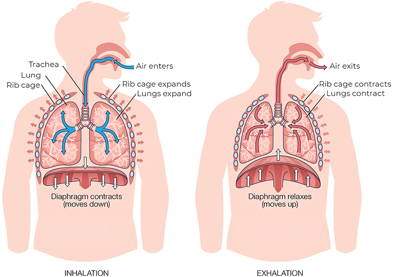
The rib cage, thoracic spine, diaphragm, abdominal wall, pelvic floor, and deep spinal stabilizers coordinate thousands of times a day to sustain respiration (Image 1). When that pattern works well, respiration supports posture, spinal stability, nervous system regulation, and fluid movement.
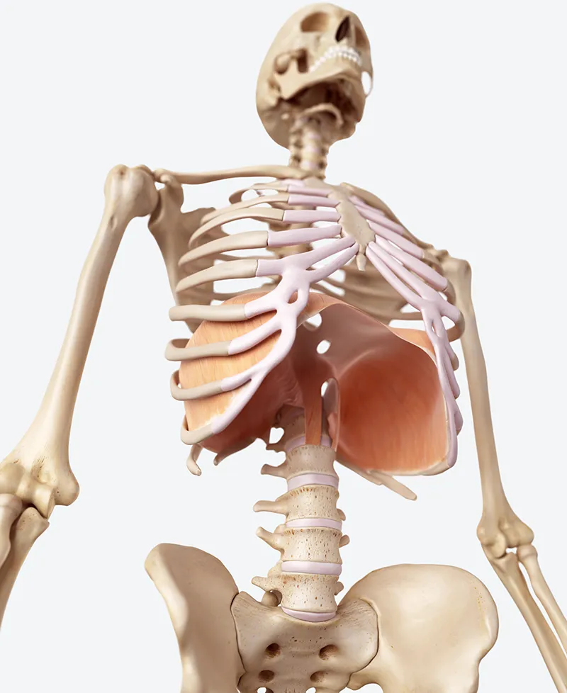
At the center of it is the diaphragm (Image 2), the primary muscle of breathing. It’s a dome-shaped sheet that separates the thoracic and abdominal cavities, attaching to the lower ribs and costal cartilages, the xiphoid process, and, through its crura, the lumbar spine. Those attachments matter to us as therapists because they link the diaphragm directly to the rib cage, the low back, the psoas, and the abdominal wall.
On inhalation, the diaphragm contracts and descends, drawing air into the lungs while the lower ribs widen. On a relaxed exhalation, it simply lets go—the diaphragm rises, the ribs recoil, and air leaves with no muscular effort (Image 1). When more force is needed, the abdominals draw the ribs down, increasing intra-abdominal pressure. That smooth descend-and-release is what we’re looking for, and it’s exactly what’s missing in the client from our introduction, whose lower ribs barely moved on the exhale.
The pressure system just described is where breathing becomes a postural matter. The diaphragm works with the abdominal wall, pelvic floor, and deep spinal stabilizers to regulate intra-abdominal pressure. We can think of this as the internal bracing that stiffens and protects the spine during lifting, reaching, walking, and rotation. When the breath coordinates well, that bracing happens automatically. When it doesn’t, the body braces inefficiently and recruits muscles that were never meant to carry the load.
Two of those connections are worth keeping in mind on the therapy table. First, the diaphragm is innervated by the phrenic nerve, which arises from C3 through C5, explaining why chronic neck tension and disordered breathing often occur together. Irritation or dysfunction in this region affects both the diaphragm’s nerve supply and the nerves serving several muscles of the neck and shoulder girdle. When the diaphragm underperforms, the body turns to the scalenes, SCM, upper trapezius, levator scapulae, and pectoralis minor for help, leaving them overburdened and hypertonic.
Second, the scapula glides across the posterior thorax through the scapulothoracic articulation, and its resting position follows the curve of the rib cage beneath it. When the thoracic spine is fixed in flexion, the scapula starts from a position of protraction, anterior tilt, and reduced upward rotation before the arm even moves.
Reaching overhead requires the serratus anterior and trapezius (especially the lower fibers) to rotate the scapula upward and tilt it backward on the rib cage. These motions depend on a mobile thoracic spine and freely expanding upper ribs. Take away that mobility and the scapula can’t rotate or tilt fully, scapulohumeral rhythm breaks down, and the cervicothoracic junction and upper trapezius take on the load.
Dysfunctional breathing rarely indicates a pathology. Most clients breathe poorly due to posture, stress, pain, habit, thoracic rigidity, or a simple lack of sensory awareness. Any single breath looks unremarkable, but repeated thousands of times a day, small inefficiencies compound into notable musculoskeletal consequences.
The most common pattern is the one our case-study client demonstrated: upper-chest, or apical, breathing. Air is drawn in high in the thorax with little lateral expansion through the lower ribs. The sternum, clavicles, and shoulders rise on inhalation while the lower rib cage stays quiet, so the scalenes, SCM, upper trapezius, levator scapulae, and pectoralis minor take over the diaphragm’s work. Over months, these accessory muscles become hypertonic and tender, producing the familiar cluster of neck tightness, jaw tension, headaches, and a sense of being wound up. The pattern travels with forward-head posture, rounded shoulders, and increased thoracic kyphosis.
A related reversal is paradoxical breathing. Normally, the abdominal wall moves outward on inhalation as the diaphragm descends. In paradoxical breathing, the abdomen pulls inward on the inhale and pushes outward on the exhale. It shows up with high stress, chronic abdominal bracing, or rib fixation, and tends to pair with rigid lower ribs and poor coordination between the diaphragm, rib cage, and abdominal wall.
Breath-holding is another common compensation. Many clients unconsciously stop breathing through the small efforts and stresses of daily life, such as lifting a box, concentrating at the keyboard, or bracing through a moment of pain or worry. Each hold raises global muscle tone and reinforces guarding. Repeated all day, it keeps the nervous system wound tighter than it should be.
This occurs when the costovertebral joints, intercostal muscles, thoracic paraspinals, or sternocostal tissues lose mobility, and the diaphragm is forced to work against a stiff container. This is why cuing a client to “breathe into the belly” sometimes fails. The rib cage lacks the capacity to expand, so the nervous system resorts to the only strategy left: the neck muscles and shoulder girdle.
Stress physiology overlays everything above. Persistent sympathetic arousal produces faster, shallower breathing, as though the body is braced for action. The upper chest lifts, the jaw tightens, the suboccipitals shorten, and the shoulder girdle guards. Each feeds the other in a cycle that reinforces itself. The tense body shapes the breath, and the shallow breath keeps the body tense, around and around, long after the original stressor has passed.
Myoskeletal alignment technique (MAT) takes a holistic approach to dysfunctional breathing, addressing the diaphragm and the structures that surround and support it. The most productive areas to address include the following.
The four related techniques that follow target restricted movement in the rib cage, the lateral abdominal wall, the diaphragm attachments, and the intercostal tissues. They are especially useful when a client is stuck in shallow upper-chest breathing and can’t easily expand the lower rib cage during inhalation.
Dalton’s thoracic and respiratory work is extensive, so the goal here is to incorporate these effective techniques into your routines to support session outcomes for clients whose neck, shoulder, and back complaints are driven by their breathing.
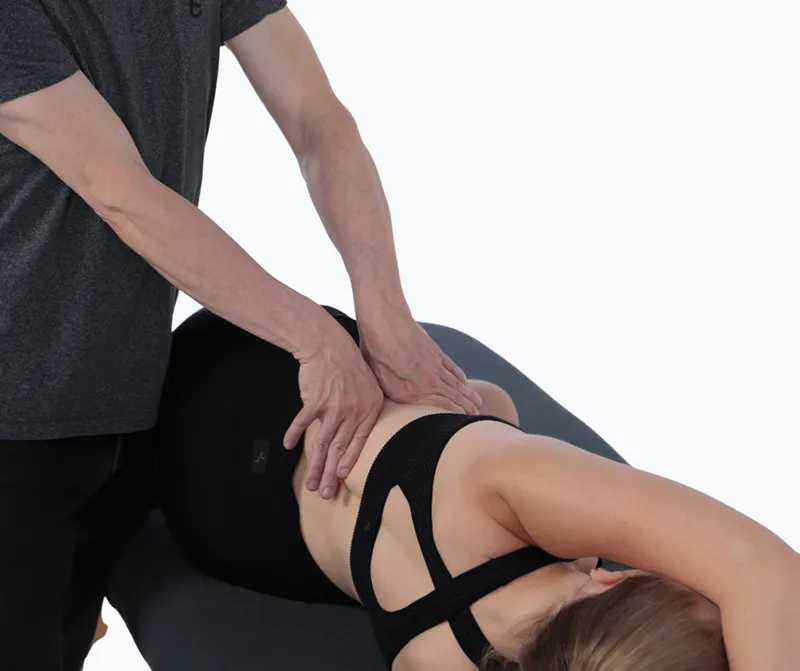
With the client lying on their left side, stand behind them at their hips (Image 3).
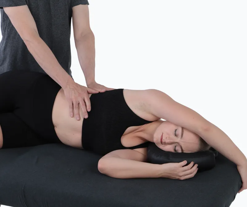
Ask the client to grasp the top of the massage table with their right hand to lengthen the lateral body. Bring your hands together and place them over the lower ribs. Ask the client to take a slow, deep breath. As the client exhales, gently drop your weight into their body and use gentle rhythmic compressions to mobilize the ribs.
Repeat the pattern for several slow breaths. Use this technique on both sides of the body.
This technique works along the costal margin to release the diaphragm’s peripheral attachments (Image 4). With the client lying on their right side, stand behind them at their hips.
Ask the client to grasp the top of the massage table with their left hand to lengthen the lateral body. With your hands held in a “V” shape, place the knuckles of your index fingers just below the ribs. Let your weight fall gently through your knuckles as the hands separate anteriorly and posteriorly. Slowly sweep your knuckles along the bottom edge of the rib cage, working with the client’s slow, deep breaths.
After the first pass, repeat the process while the client performs slow pelvic tilts and continues to take slow, deep breaths. Repeat the technique several times on both sides of the body.
This time, practice with the client lying on their left side (follow along in the correlating video). Have the client lie close to the edge of the massage table (Image 5). Their left leg is bent, and their right leg is straight. The client places their right hand behind their head. Place your right hand along the costal margin of the client’s ribs with your fingers on the anterior side, contacting the transabdominal fascia. Your left hand contacts the client’s right elbow.
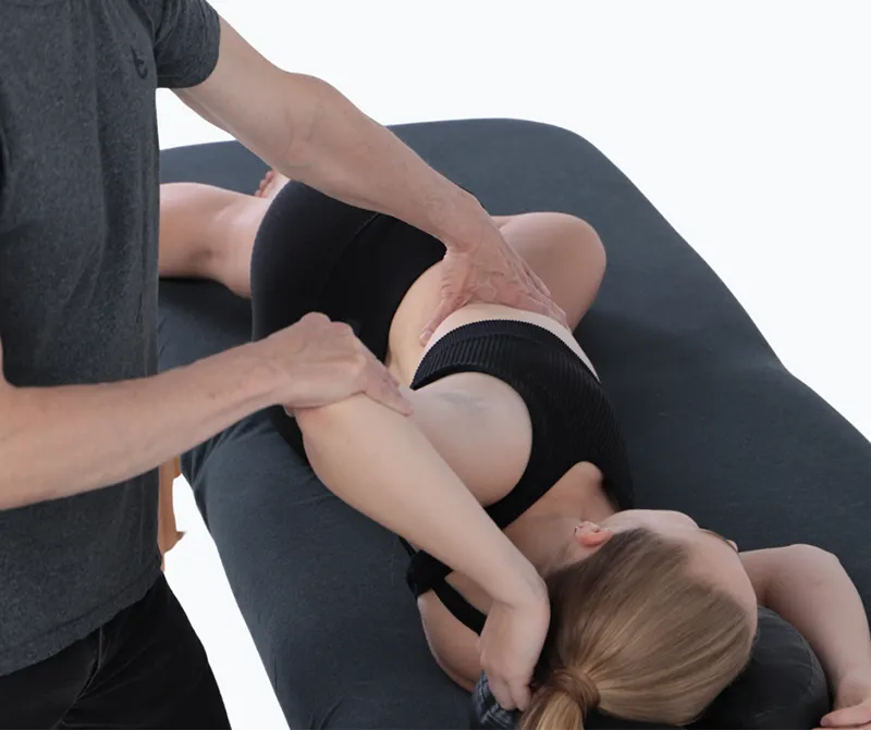
Ask the client to press their elbow forward into your resistance as they inhale, then hold for five seconds. As the client exhales, use the thumb and fingers of your right hand to firmly pin the client’s tissue along the costal margin while your left hand uses the elbow to stretch and open the client’s body by pulling the elbow gently backward. Repeat this technique several times on both sides of the body.
The client is lying on their left side, close to the edge of the massage table (Image 6). Their left leg is bent, and their right leg is straight (as shown in the second half of the video). The client’s right arm is extended over the top of the therapy table. Turn your back to the client and anchor the hip with your left hand, leaning back slightly. Press your right hand up under the costal margin of the ribs, to pin the tissue.
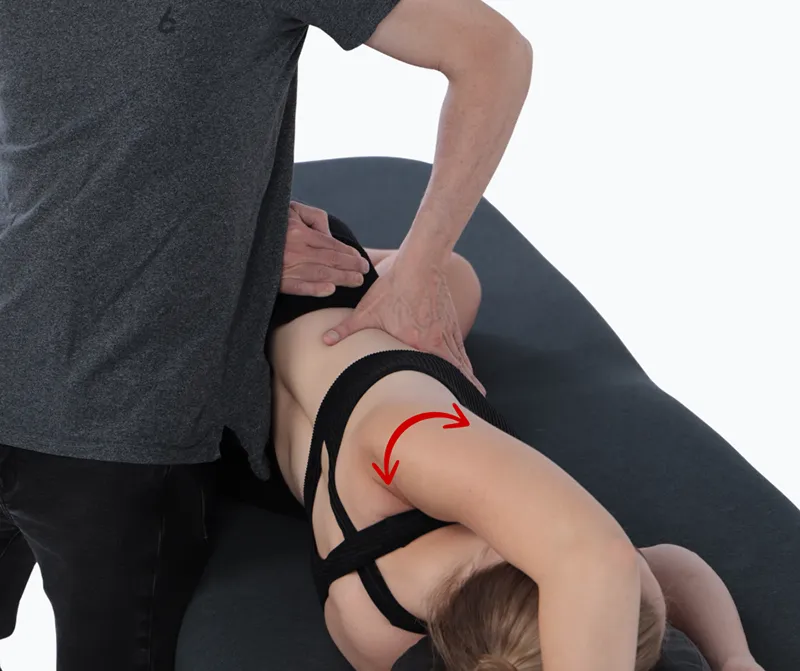
Ask the client to inhale and exhale slowly while they externally and internally rotate their arm at the shoulder joint. When the client inhales, they externally rotate the arm; when they exhale, they internally rotate the arm as you take out the slack in the tissue with your right hand. Repeat this technique several times on both sides of the body.
Breathing is the movement pattern our clients repeat more than any other, and it’s the one most often overlooked in the treatment room. The techniques we shared here offer a way to address the pattern underlying many of the complaints you already treat. If respiration is our primary and most dysfunctional movement pattern, then techniques to free our breathing are central to achieving optimal client outcomes.
Just because the pain is in the neck doesn’t mean that’s where it’s originating.
To understand tissue-tension barriers, MTs must be constantly present and mindful. Too light and we will not become efficient at creating the healing change that is needed.
New research into hip pain can provide you with more options for supporting clients in that region.
“Energy” or “life force” can have many definitions. As we treat clients, we should consider how its various meanings can be interpreted and applied to our work.