Historically, when clients come in complaining of a persistent, nagging ache on the outside of the hip, the pain is attributed to inflammatory “trochanteric bursitis,” an inflamed bursa. Treatment focused on local anti-inflammatories or corticosteroid injections. However, new research has given us a better understanding of the structural reasons for this often-chronic pain syndrome. For the massage therapist, these fresh insights require us to adapt our care.
Similar to the reconsiderations of tendinitis, lateral hip pain also appears to be less about inflammation and more about tendon degeneration and compression mechanics. Three common conditions can be implicated in lateral hip pain: gluteal tendinopathy, trochanteric bursitis (in 8 percent of cases), and a third, less common condition called snapping hip syndrome (the popping or clicking sensation some feel or hear when standing up from sitting or walking that happens when the tendon or muscle “snaps” over the greater trochanter). The umbrella term for these three conditions is greater trochanteric pain syndrome (GTPS).
GTPS is a widespread issue, affecting an estimated 10 to 25 percent of the population in developed countries.1 GTPS primarily affects middle-aged and elderly adults, with women being affected four times more often than men. GTPS is frequently chronic and sometimes lasts several years. It can become a serious disability, on the level of end-stage hip osteoarthritis. Let’s look at how these new insights change our approach to treating lateral hip pain.
Anatomy and Biomechanics
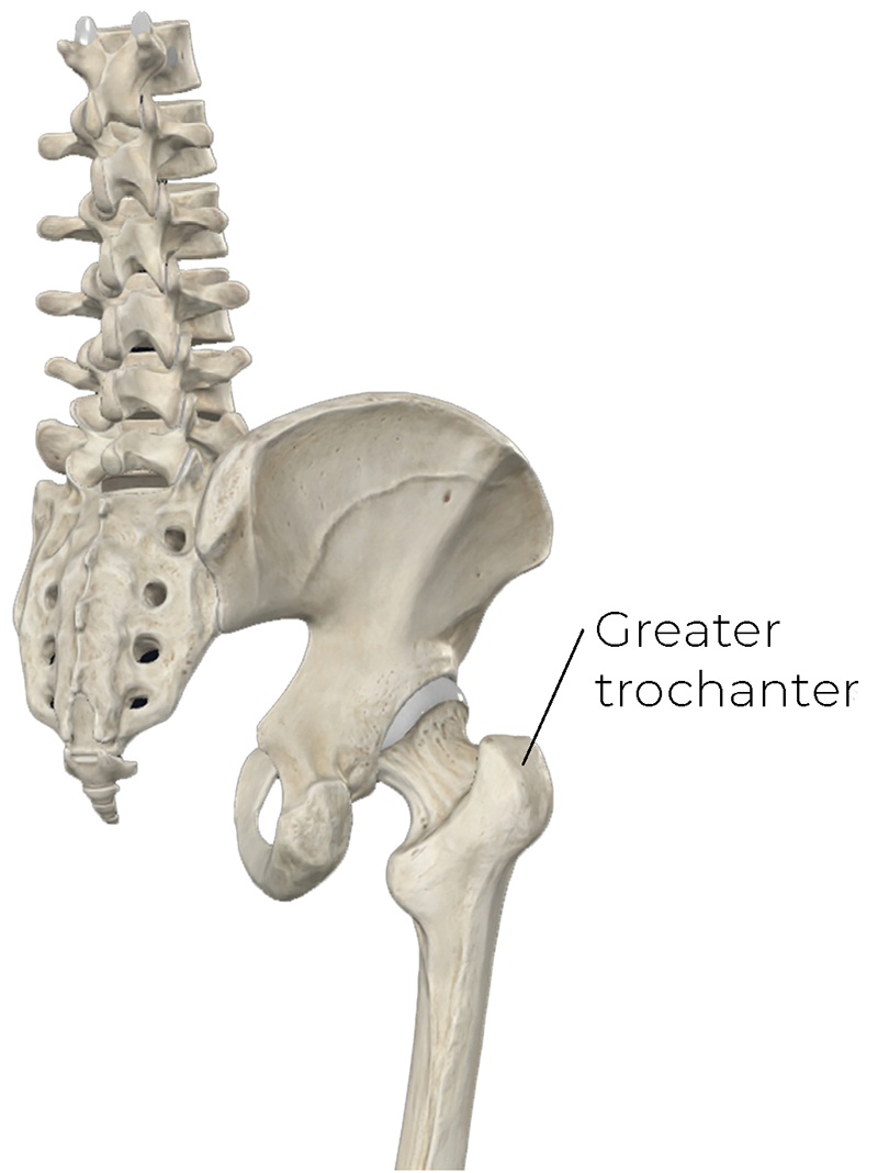
To build an effective treatment plan, we need to understand the anatomy and biomechanics in the lateral hip region. The greater trochanter of the femur is the large bony projection that is easily palpable on the lateral hip (Image 1). This projection serves as a primary attachment site for the hip abductors and the hub for the large forces generated by these muscles.
Image 1. The greater trochanter of the femur. Image courtesy Complete Anatomy.
The Abductor Mechanism
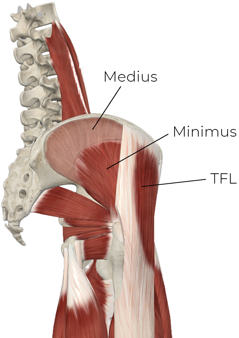
Lateral hip stability is achieved through the abductor mechanism, which includes the gluteus medius and gluteus minimus muscles, and is supported superficially by the tensor fasciae latae (TFL) (Image 2). Together, these muscles are essential for maintaining pelvic stability and preventing the pelvis from dropping on the opposite side during the stance phase of gait.
Image 2. The hip abductors and tensor fasciae latae (TFL). Image courtesy Complete Anatomy.
Recognizing the relationship among these deep tendons and muscles, the overlying iliotibial (IT) band, and the local bursa is critical to understanding GTPS. The IT band effectively functions as a stiff, compressive “roof” over the underlying gluteal tendons and muscles. Structural compression by the overlying IT band is a major cause of pain and tissue degeneration in GTPS.
Pathophysiology: From Tensile Strain to Compressive Loading
In GTPS, current understanding leads us away from tendinitis and inflammation toward tendinopathy, though trochanteric bursitis remains a possible cause of pain. However, this pain syndrome overwhelmingly involves degeneration and failed healing of the hip abductor tendons and the gluteus medius and gluteus minimus. This degenerative state is characterized by cellular changes, including collagen matrix disorganization and neovascularization (excess blood vessels and small-fiber nerve endings).
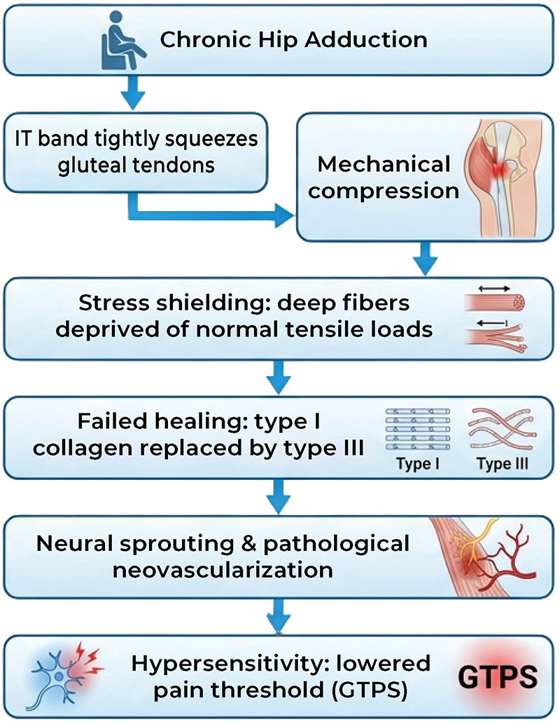
A cellular change occurs in affected tendons in which healthy Type I collagen is replaced with structurally weaker Type III. Type I collagen is thick, with parallel and highly cross-linked fibers. The body produces Type III collagen to act as a quick fix. It has a more mesh-like, disorganized fiber pattern that is thinner and weaker. Type I resists tension and stretching; Type III is more elastic and flexible. A common comparison is that Type III is more like a rubber band, while Type I is like a leather strap. You can see a flowchart for this process in Image 3.
Image 3. Greater trochanteric pain syndrome and the pathway of tendon degeneration. Image courtesy Whitney Lowe.
While tendons are structurally designed to resist tensile pulling forces, they adapt poorly to compressive loads. In the hip, compression peaks during adduction, when the IT band is pulled taut over the underlying fibers of the gluteus medius and minimus tendons, thereby forcing them against the rigid, bony greater trochanter.
Chronic compression triggers a phenomenon known as stress shielding, which occurs when a tendon is squeezed so tightly against a bone that the tendon is prevented from experiencing the normal pulling forces it needs to remain strong and healthy. Without this normal mechanical stretch, the tendon essentially wastes away, becoming fragile and a significant source of chronic pain.
As the body attempts to repair the degraded tendon, pathological neovascularization occurs. Unfortunately, this abnormal blood vessel growth is accompanied by neural sprouting—the ingrowth of small, nociceptive nerve fibers. These new nerve endings lower the local pain threshold and are a primary source of the pain associated with tendon degeneration.
Biological risk factors accelerate this pathology. For example, postmenopausal declines in estrogen are linked to tendon pathology. Concurrently, those with wider pelvises will experience naturally elevated IT band mechanical compressive force over the greater trochanter, explaining why women suffer from this condition more than men.
Assessment Principles
When a client presents with lateral hip pain, our key therapeutic goals are to track tissue irritability, identify contributing structures, and check for red flags. Assessment begins with information from the client history.
The pain behavior of GTPS is highly characteristic. It’s localized to the lateral hip and frequently starts as an intermittent, dull ache that gradually intensifies during weight-bearing activities, prolonged walking, climbing stairs, or lying on that side.
A classic indicator is night pain. Clients consistently report severe sleep disruptions caused by direct mechanical compression when lying on the affected side. Conversely, lying on the unaffected side can be equally painful if the top (injured) leg crosses over the midline of the body into hip adduction. This adducted position pulls the IT band taut, wrapping it tightly over the greater trochanter and squeezing the underlying tissues.
Physical Exam Findings
A series of physical tests that simulate the tensile and compressive forces likely to reproduce the client’s primary pain can be used to confirm our clinical suspicions:
Focal palpation—One highly sensitive test is to apply direct pressure just above (superior to) the greater trochanter. Palpating the tendon during resisted hip abduction is even more likely to reproduce symptoms. The objective here is not to aggravate symptoms but rather to confirm the most likely cause of the client’s discomfort.
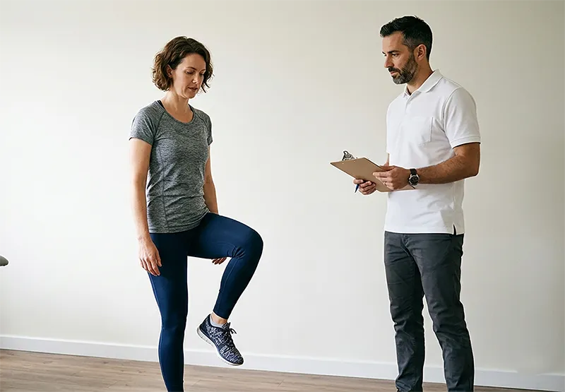
30-second single-leg stance (SLS) test—This is a high-specificity test in which the client is instructed to stand unsupported on the affected limb. The reproduction of lateral hip pain within 30 seconds signifies a positive test, pointing to an inability of the abductor mechanism to handle sustained tensile and compressive loading (Image 4).
Resisted hip abduction—This test isolates contraction-induced tensile loads. To test, place a hand on the distal, lateral femur just above the knee and have the client attempt to further abduct the hip against your resistance. This tests for weakness, tendon degradation, or pain inhibition.
Differentiating GTPS from hip osteoarthritis (OA)—It’s critical to differentiate regional GTPS from intra-articular hip OA. Unlike clients with hip OA, those with GTPS generally retain the full range of motion needed to bend down and put on their shoes and socks. Those with OA tend to lose range of motion and have difficulty getting into this position. GTPS pain presents distinctly on the lateral aspect of the hip, whereas OA pain presents deep within the anterior groin.
Image 4. The single-leg stance test. Image courtesy Whitney Lowe.
Key Treatment Strategies
When addressing GTPS, manual therapy plays a vital, strategic role in the early stages of recovery. Targeted massage is highly effective for reducing muscular tightness, easing local guarding, and calming nervous system hyperreactivity. It’s also highly beneficial for pain management. Research suggests that massaging affected tendons may stimulate fibroblast proliferation, thereby aiding the healing process.
By reducing hypertonicity and managing short-term pain, we effectively prepare the hip region for rehabilitation exercises. Initial massage treatment provides a transition period for the progressive tendon loading that occurs during physical therapy exercise. Tendon loading is necessary for tissue regeneration and encouraging Type I collagen remodeling.
Addressing the “IT Band Roof”
To decompress the affected tendons, we must treat the muscular structures that govern the tension of the IT band. Because the massive gluteus maximus muscle and TFL both insert directly into the IT band, chronic hypertonicity in these muscles dramatically increases down-bearing, lateral compressive tension over the greater trochanter. Manual work should focus on myofascial and trigger-point therapy applied directly to the TFL and the gluteus maximus. Massage with active movement is particularly helpful for reducing hypertonicity and enhancing pain management.
It’s also important to evaluate and treat muscle guarding in the hip adductors and lumbar muscles. These secondary muscle groups routinely overwork to stabilize the pelvis during gait when the primary gluteus medius has become inhibited by pain.
Technique Guidelines
When applying specific manual techniques, client positioning and appropriate technique are key to achieving beneficial outcomes. When working near the abductor tendon insertions, avoid compressing them directly against the bone while they are taut. To minimize bursal friction and properly isolate these deep structures, position the client supine with the hip passively abducted to 45 degrees. This position relaxes the overlying IT band, allowing you to gently access the deep insertions without grinding the tendons against the trochanter.
Begin with more superficial myofascial applications. Employ sustained, broad pressure to reduce protective fascial guarding across the lateral hip and thigh. Myofascial applications (especially those with a broad contact surface) improve multidirectional tissue glide, ensuring the IT band slides smoothly over the trochanteric region.
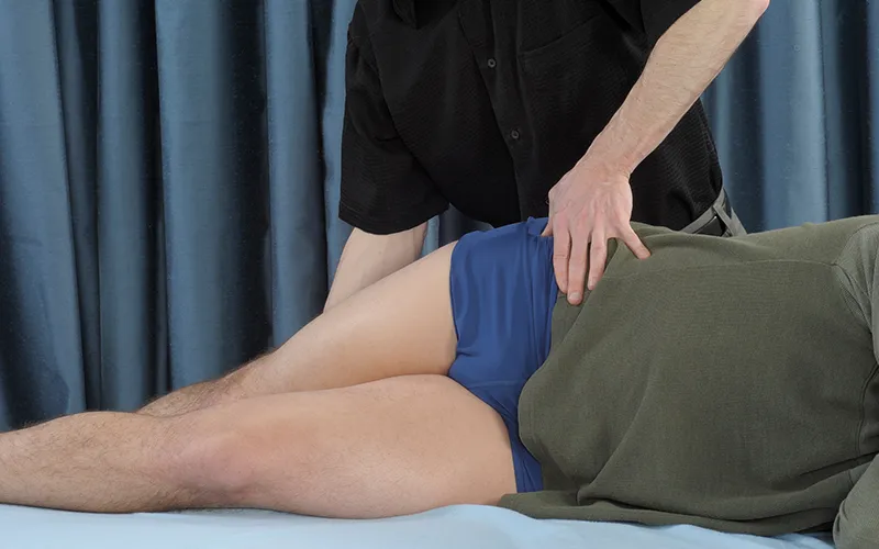
Next, apply direct manual pressure with a small contact surface or use stripping techniques to the hip abductor muscle bellies during active eccentric elongation (Image 5). This treatment will enhance fascial mobility through mechanotransduction, the process by which physical forces—such as massage pressure and muscle stretching—signal cells to repair, adapt, and restructure tissue, thereby improving mobility and elasticity.
Image 5. Compression or stripping during eccentric hip adduction. Image courtesy Whitney Lowe.
Simultaneously, this therapy stimulates sensory cells via the gating mechanism, producing a multilayered effect valuable for safely working through deep, thick muscle groups like the hip abductors and gluteal muscles.
Red Flags, Contraindications, and Precautions
Safe clinical practice respects scope-of-practice boundaries and medical red flags and recognizes when to refer.
Medical Red Flags and Referral Metrics
Watch for these medical red flags.
Femoral neck stress fractures—This is a repetitive-use bone injury of the femoral neck. It shows up as deep groin, hip, or anterior thigh pain. The pain worsens with weight-bearing activities, running, or hopping. It is pain that can be similar to the early stages of GTPS. Clients suspected of this injury should be referred to an orthopedist for imaging.
Systemic or infectious pathologies—Therapists should be suspicious if a client presents with active red flags such as localized heat over the hip, unexplained skin redness, systemic fever, or sudden, unremitting night pain that is unrelated to mechanical positioning.
Direct Contraindications
Keep these direct contraindications in mind.
Recent injections—Be sure not to apply pressure or massage over the space around the trochanter for at least 48–72 hours following a local corticosteroid or platelet-rich plasma (PRP) injection. This will protect the sterile environment and allow the injected solution to integrate without disruption.
Known unstable fractures—Massage, stretching, or manual therapy should not be applied if an underlying fracture is suspected or medically confirmed.
Clinical Precautions and Irritability Modifications
Acute irritable bursitis—When the trochanteric bursa is actively inflamed, direct deep-tissue compression over the greater trochanter is excruciatingly painful and worsens swelling. Focus instead on lighter, more proximal treatments for pain management.
Adduction intolerance—Therapists must avoid stretching the lateral hip, IT band, gluteal muscles, or piriformis into end-range hip adduction. Bringing the leg across the midline directly replicates the compressive mechanism that caused the tendinopathy.
Sidelying position strategy—Prolonged sidelying on a standard treatment table can severely aggravate GTPS due to gravity-induced compression. Therapists should position supportive pillows between the client’s knees to maintain strict neutral hip alignment or transition the client to a supine or other more comfortable position.
Clinical Tips for Therapists
Five clinical tips to keep in mind:
Stop “stretching” the lateral hip—Traditional IT band or piriformis stretching forces the hip into adduction, which tightens the fascial band across the trochanter and increases compression on the injured tendons and muscles. Focus instead on tendon loading, pain reduction, and relieving hypertonicity.
The 24-hour rule—Watch out for posttreatment soreness. If lateral hip pain increases and does not return to the client’s baseline within 24 hours, the treatment was too aggressive and must be scaled back in depth or duration during the next session.
Target the big glute—Because about 80 percent of the gluteus maximus fibers insert directly into the IT band, treating this major muscle belly is far more effective at reducing lateral band compression than massaging the dense, unyielding IT band. However, there can still be some degree of pain management and symptom reduction by working on the lateral thigh region as you affect the muscles underneath the IT band.
Hips higher than knees—Instruct clients to change how they sit; keeping the hips elevated above the knees reduces the sharp angle of hip flexion and subsequent adduction that compresses the muscle and tendons.
Address daily compression habits—For lasting recovery, help your clients become aware of potential postural stresses. Such stressors include standing with weight on the affected side and habitual leg crossing while sitting. For side sleepers, encourage them to try sleeping on their backs or to place padding between their legs if sidelying.
Guide Clients out of Pain
Manual management of GTPS requires a paradigm shift away from treating an inflammatory “bursitis” toward managing a compressive, degenerative tendinopathy. Massage therapy provides an invaluable, evidence-based service. Massage is one of the best treatments for calming a sensitized nervous system. Our therapies also work to ease secondary muscle guarding along the kinetic chain and create an optimal, pain-free window for active rehabilitation.
By combining targeted manual therapy with client education on load management and a supportive, progressive exercise program, we can guide our clients out of chronic pain and back into full, comfortable function.
To understand tissue-tension barriers, MTs must be constantly present and mindful. Too light and we will not become efficient at creating the healing change that is needed.
“Energy” or “life force” can have many definitions. As we treat clients, we should consider how its various meanings can be interpreted and applied to our work.