

When Superficial Fascia Goes Deep
Superficial fascia may not get the same attention as deep fascia, but it plays an extremely important role in the organization of tissues and the structure of the body.
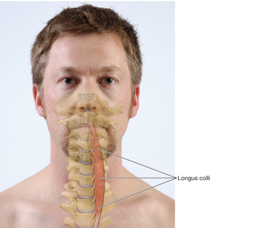
The longus colli is a deep cervical muscle located on the anterior surface of the vertebral column, deep to the thyroid, trachea, and esophagus. This relatively flat muscle spans the anterior surfaces of the vertebral bodies between the atlas (C1) and third thoracic vertebra. The deepest of the anterior neck muscles, the longus colli is long and primarily vertical with multiple segments. The superior oblique portion originates on the anterior tubercles of the transverse processes of C3-5, ascends obliquely and medially, and narrows to insert onto the anterior arch of the atlas.
A middle vertical portion originates on the anterior bodies of C5-T3, ascends vertically, and inserts on the anterior bodies of C2-4. A third inferior oblique portion originates on the anterior bodies of T1-3, ascends obliquely and laterally, then inserts on the anterior tubercles of C5-6. Together, these segments create an interconnecting network between the anterior surfaces of the cervical and upper thoracic vertebrae.
The longus colli is a strong flexor of the head and neck when both sides fire, as it spans all of the cervical vertebrae and is segmented. It is often associated with the rectus capitis anterior and rectus capitis lateralis as the paravertebral group. This group helps stabilize the anterior neck during high-intensity activities like sneezing and rapid arm movements like throwing. It also actively stabilizes the front of the curve of the neck—counteracting the lordotic curvature of the cervical spine due to the weight of the head—and keeps the head from falling back.
Longus colli is clearly divided into right and left sides with a gap at the midline of the vertebral bodies. This creates some leverage for lateral flexion. Oblique fiber orientation in its superior and inferior segments generates slight rotation to the opposite side when longus colli fires unilaterally.
Weakness in the longus colli is common, and this muscle is often affected by whiplash injuries. Poor postural control and cervical stabilization may result and contribute to chronic hypertonicity of the sternocleidomastoid and anterior scalene muscles as they attempt to compensate. In extreme cases, cervical instability and symptoms of vertigo may occur. Dysfunction of the longus colli is observed as forward-head posture with associated hypertonicity, adhesions, and trigger points in the compensating muscles. Clients also demonstrate difficulty or inability to perform segmented cervical flexion against gravity without thrusting the chin forward.
Manual techniques that address the compensatory pattern, as well as the underlying injury to the longus colli, help restore function. In some cases, referral for neuromuscular retraining and therapeutic exercise may be necessary for maximal recovery.
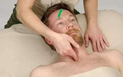
Positioning: client supine.

Editor's note: The Client Homework element in Functional Anatomy is intended as a take-home resource for clients experiencing issues with the profiled muscle. The stretches identified in Functional Anatomy should not be performed within massage sessions or progressed by massage therapists, in order to comply with state laws and maintain scope of practice.
Liu, Xiao-Ming et al. "Does the Longus Colli Have an Effect on Cervical Vertigo? A Retrospective Study of 116 Patients." Medicine 96, no. 12 (2017): e6365.
Physiopedia. "Deep Neck Flexor Stabilisation Protocol." Accessed July 2017. www.physio-pedia.com/Deep_Neck_Flexor_Stabilisation_Protocol.
Superficial fascia may not get the same attention as deep fascia, but it plays an extremely important role in the organization of tissues and the structure of the body.
Understanding tendons—their shapes, lengths, and organization—improves an MT’s touch vocabulary and facilitates a more skilled touch.
While the neck is a bridge, a pathway, the position of the neck and head can also indicate a multitude of other things happening beneath the surface.
Understanding fibroblasts and the extracellular matrix changes how we think about the tissue we touch.