

Fascia: The Pericardium-Diaphragm Connection
Understanding how the pericardium relates to breathing can in turn help clients relax into their massage session.
Continuing our examination of proprioception that began in the Massage & Bodywork March/April 2016 issue (“Muscle Spindles”), we will now examine the role of articular mechanoreceptors. These sensory structures reside around and within synovial joints and use mechanical deformation to register and report joint position, movement direction, and movement speed.
The information gathered by joint mechanoreceptors tends to be subtler than that of their muscular counterparts, as these structures are activated by relatively small amounts of stimulus (low threshold detection). Despite this, the information they provide has significant influence over neuromuscular control mechanisms, influencing the activity of the entire proprioceptive system at multiple segments in the kinetic chain.
The three types of articular mechanoreceptors are:
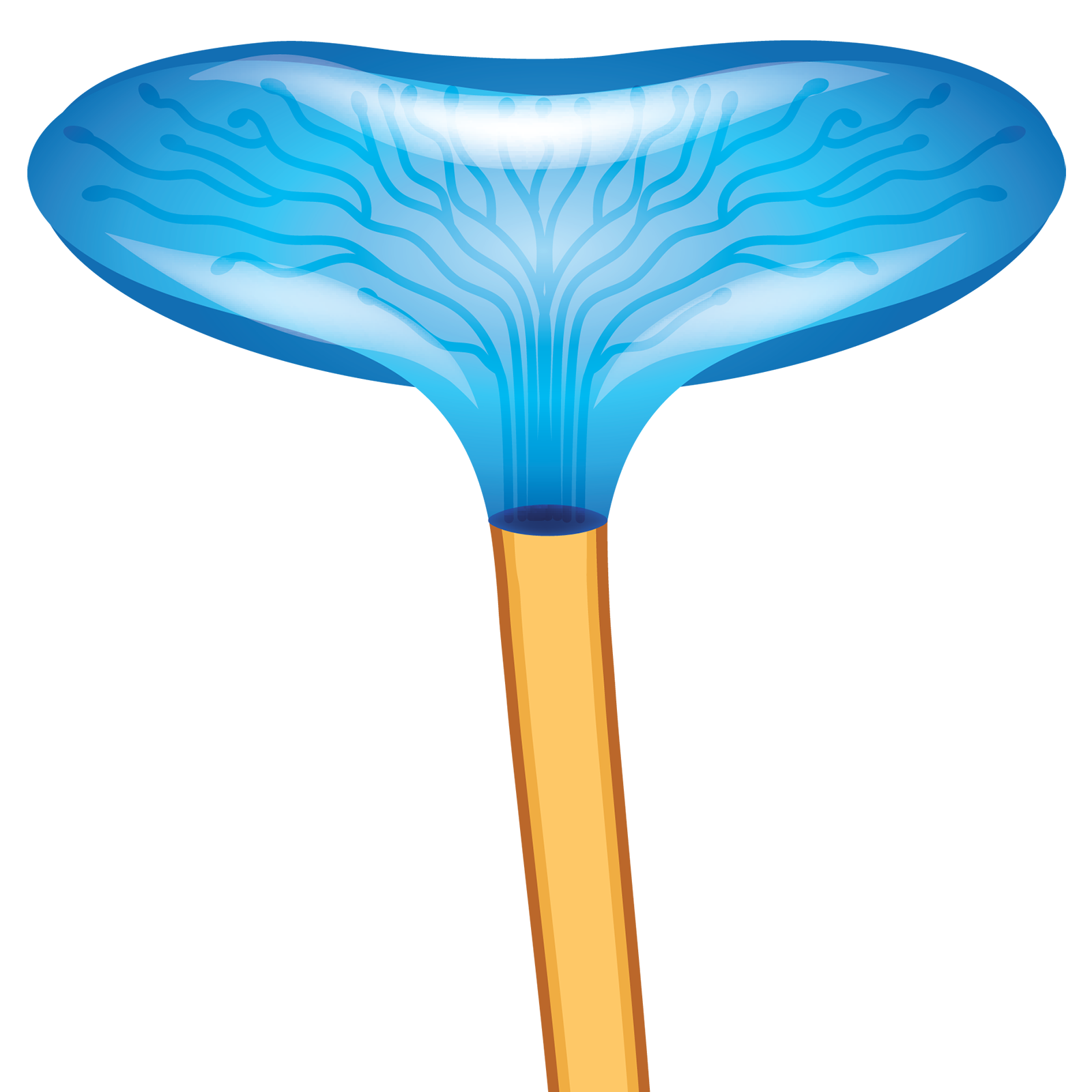
Type I, Ruffinian—Globular or ovoid corpuscle with a thin capsule.
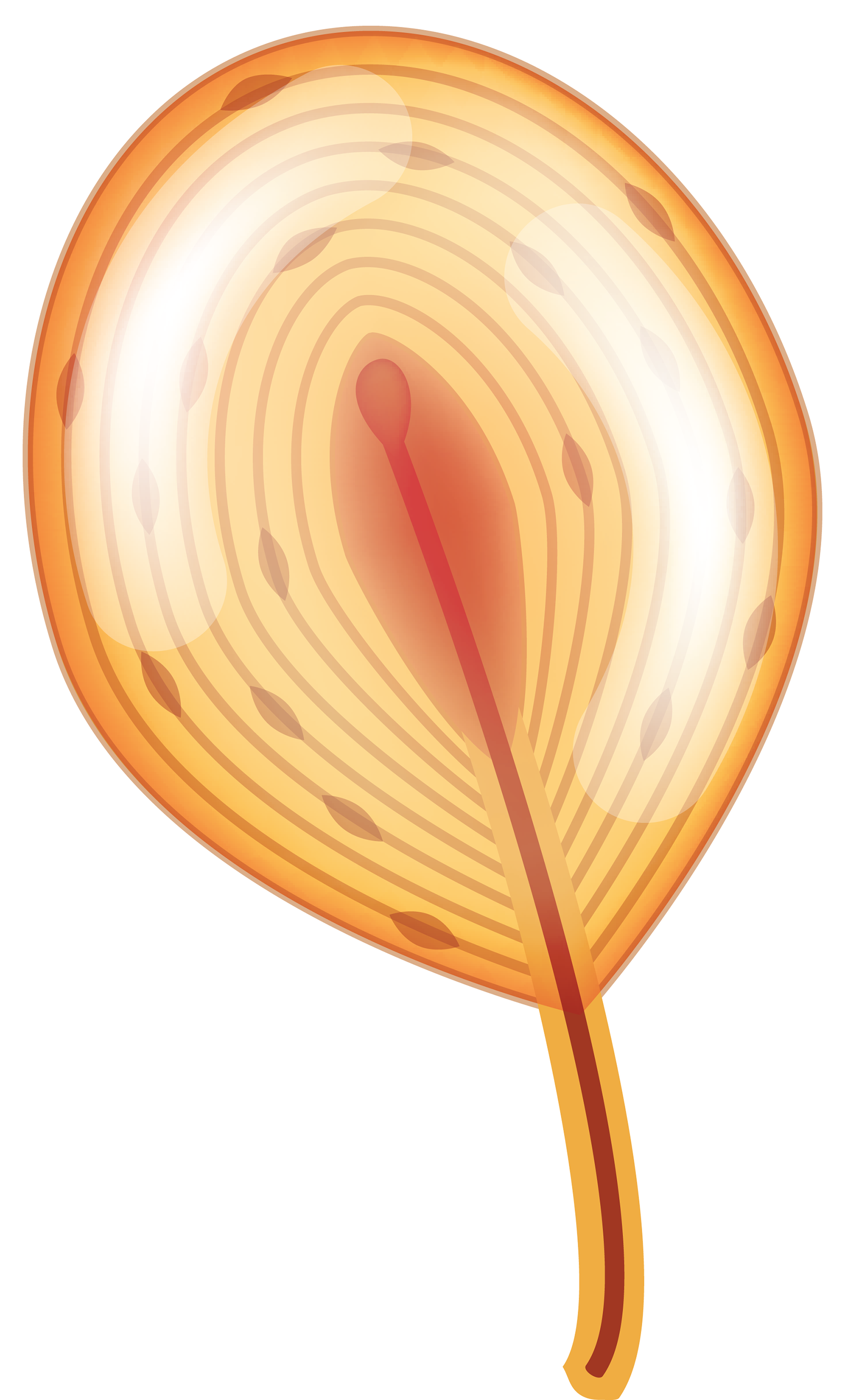
Type II, Pacinian—Cylindrical or conical corpuscle with thick, lamellated, or plated capsule.
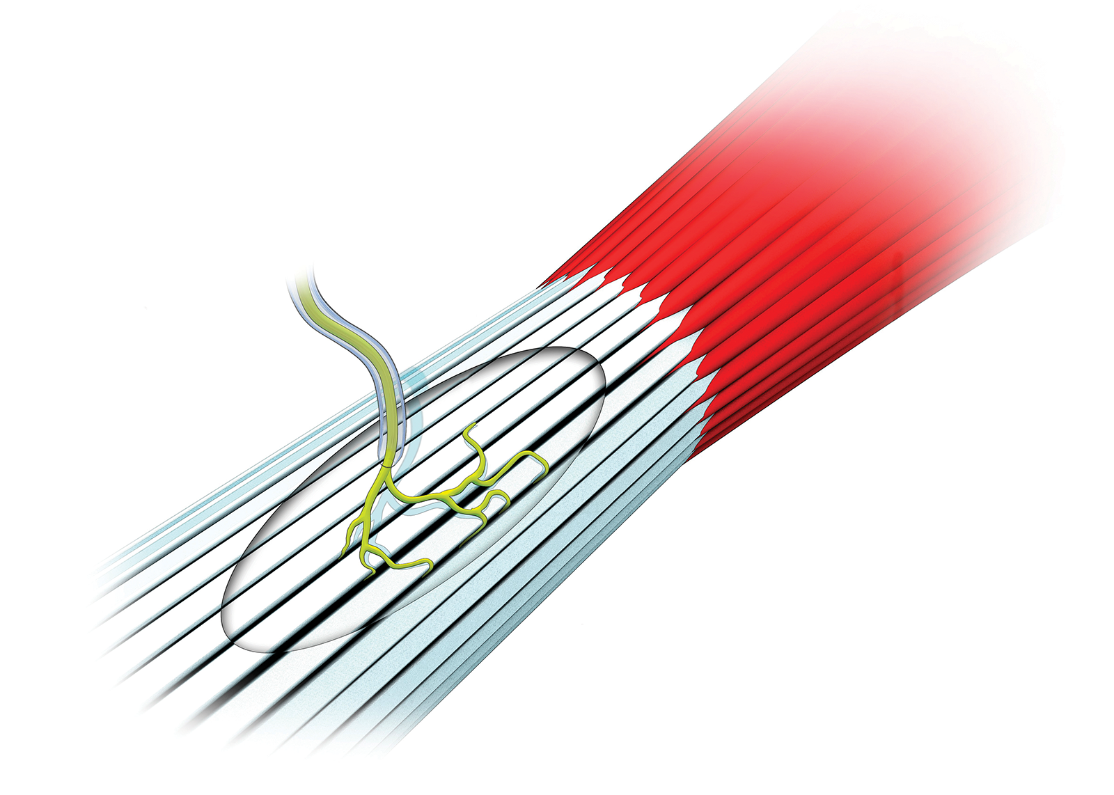
Type III, Golgi-like—Fusiform corpuscle with thin capsule.
Structurally, all of these articular mechanoreceptors include a distinctly shaped body that contains a sensory nerve and is surrounded by an outer covering or capsule. As joint movement occurs, the shape of the corpuscle is distorted via compression, tension, or shear forces. Imagine squishing a rubber ball. With enough distortion, a signal is initiated in the sensory nerve relaying the size, speed, and direction of joint motion. Imagine covering the floor with rubber balls and squishing a series of balls in a given order at a given speed. The signals of individual mechanoreceptors are relatively small and difficult to interpret, but collectively they provide tremendous amounts of proprioceptive information.
Understanding how the activity of articular mechanoreceptors influences movement starts with what is happening outside the body, or external stimulus. Vision and proprioception work in tandem to predict necessary movement patterns and make adjustments as those movements are executed. For example, visual input is combined with joint and muscle proprioceptive input to recognize uneven ground when hiking. Specific movement patterns are selected to navigate the surface and corrections are made as the patterns are executed based on both visual and proprioceptive cues. This process relies on information that is both feed forward (changes predicted based on past experience) and feedback (changes detected and adjustments made).
The body must also respond to changes inside the body, or internal stimulus. As movements are executed and adjusted, changes in joint position lead to altered leverage or mechanical advantage for all involved muscles. Force production amounts and directions must be adjusted based on these changing conditions. You are able to maintain a steady pace and rhythm even though your joints are moving through a greater range of motion as the trail becomes steeper.
If everything is working properly, articular mechanoreceptors help coordinate specific patterns of muscular facilitation and inhibition at multiple joints and segments of the kinetic chain, ending in a smooth, coordinated movement pattern. If not, inefficient joint mechanics, postural deviations, and compensatory movement strategies appear. Additionally, associated free nerve endings are no longer inhibited, increasing pain signals in these same regions. Bodyworkers help clients maintain or restore articular mechanoreceptor function following injury by reducing excessive joint compression, encouraging proper joint alignment, and restoring full, pain-free active and passive range of motion.
Understanding how the pericardium relates to breathing can in turn help clients relax into their massage session.
Superficial fascia may not get the same attention as deep fascia, but it plays an extremely important role in the organization of tissues and the structure of the body.
Understanding tendons—their shapes, lengths, and organization—improves an MT’s touch vocabulary and facilitates a more skilled touch.
While the neck is a bridge, a pathway, the position of the neck and head can also indicate a multitude of other things happening beneath the surface.