

Fascia: The Pericardium-Diaphragm Connection
Understanding how the pericardium relates to breathing can in turn help clients relax into their massage session.
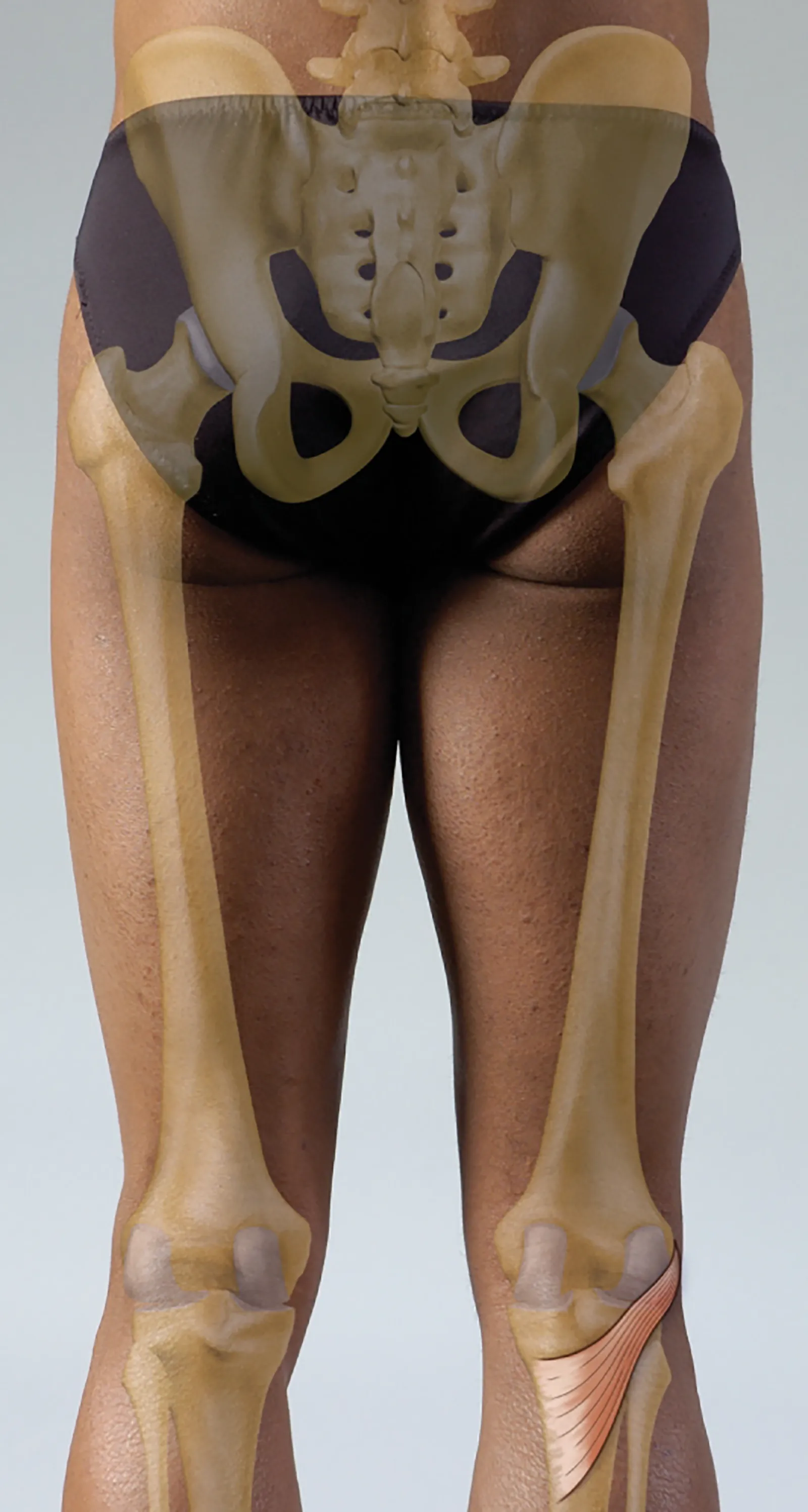
The triangular-shaped popliteus muscle is located at the back of the knee and forms the floor of the popliteal fossa. The popliteal fossa is a shallow depression outlined medially by the semimembranosis and semitendinosis tendons and medial head of the gastrocnemius, and laterally by the biceps femoris and lateral head of the gastrocnemius. This depression contains several essential structures, including the tibial nerve, common peroneal nerve, popliteal artery and vein, small saphenous vein, and popliteal lymph nodes and vessels. Caution should be exercised when locating the popliteus muscle within the popliteal fossa to avoid compressing these structures.
The fibers of the popliteus originate on the lateral condyle of the femur, run deep to the arcuate popliteal and lateral collateral ligaments, and then spread distally and medially to a broad attachment on the posterior tibia. The oblique orientation of the muscle creates rotation at the tibiofemoral joint in addition to flexion of the knee. If the foot is planted or fixed when the popliteus is activated, the femur will rotate laterally on the tibia. If the foot is not fixed, the tibia will rotate medially on the femur.
The main function of the popliteus muscle is to unlock the locked, fully extended knee. This “screw-home” mechanism is possible because the medial femoral condyle is larger than the lateral, allowing slight rotation of the knee at end-range extension. As the knee fully extends, the tibia spins externally on the femur until it reaches full external rotation. This is the “locked” position. Locking the knee decreases the work of the quadriceps muscles when standing.
A simple way to observe the rotational motion of the screw-home mechanism is to sit in a chair facing forward. Straighten one knee fully, watching the position of your foot as you do so. As you reach full knee extension, your foot should turn out slightly. This happens when the tibia turns on the femur. You can also observe it by standing and gently locking (fully extending) and unlocking (slightly flexing or softening) the knee.
Hyperextension of the knee can damage the popliteus muscle, creating pain and swelling in the back of the knee, as well as dysfunction in the lower extremity. The popliteus is also vulnerable to injury when planting and pivoting around the lower extremity. Weakness or inhibition of the hamstring muscles, particularly with rotational movements, may contribute to excessive stress, pain, trigger point activation, and dysfunction in the popliteus muscle.
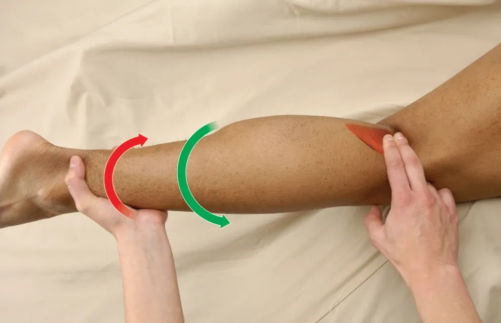
Position: client prone with knee slightly flexed.
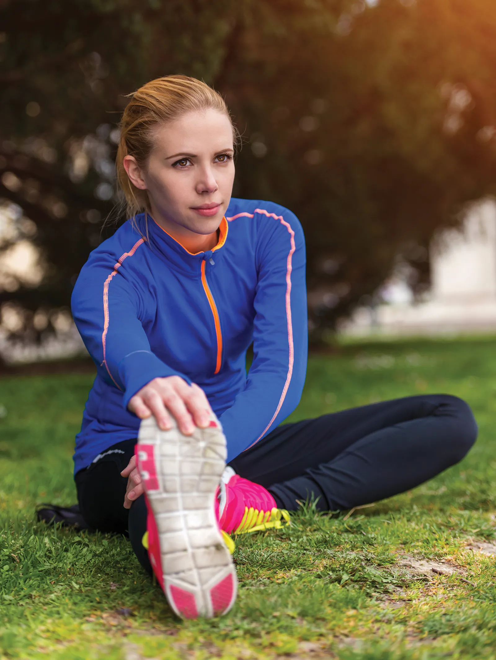
Editor’s note: The Client Homework element in Functional Anatomy is intended as a take-home resource for clients experiencing issues with the profiled muscle. The stretches identified in Functional Anatomy should not be performed within massage sessions or progressed by massage therapists, in order to comply with state laws and maintain scope of practice.
Understanding how the pericardium relates to breathing can in turn help clients relax into their massage session.
Superficial fascia may not get the same attention as deep fascia, but it plays an extremely important role in the organization of tissues and the structure of the body.
Understanding tendons—their shapes, lengths, and organization—improves an MT’s touch vocabulary and facilitates a more skilled touch.
While the neck is a bridge, a pathway, the position of the neck and head can also indicate a multitude of other things happening beneath the surface.