


When Superficial Fascia Goes Deep
Superficial fascia may not get the same attention as deep fascia, but it plays an extremely important role in the organization of tissues and the structure of the body.
In every massage we perform, whether supine, prone, sidelying, or semi-reclining, how often do we consider what’s happening behind muscles and ribs, in the land of the internal organs? How are they organized in there? What holds them in place? Are they connected to each other? We are so familiar with the sensations of our internal connections that we take them for granted.
For example, let’s start with a quick sensory experiment. Take a full breath and notice what moves. Did you feel your chest expand and then settle? What about your ribs? Sternum? Collarbones? Maybe your belly rose and fell too. Now imagine taking a deep breath and not feeling any connected movement through your chest and torso. It seems impossible. Breathing makes things move.
Next, tune into your heartbeat. Notice how subtly you can feel it, if at all. Now, think about what it would be like if every heartbeat pulled your sternum and ribs down with a strong twist. It would be weird, right?
Our breath naturally moves elements of our torso as the diaphragm contracts, but how? What is it connected to? And when our heart beats, we can feel our pulses, of course, but the beating doesn’t create pulling in our chest. Why? Let’s look inside the trunk and examine the function and relationship between the heart, pericardium, and diaphragm—parts we don’t directly touch with our massage but that can impact how well we move, both inside and out.
Every one of your organs has a connective tissue wrapping around it that is a part of the fascial system: the visceral fascia. This category of fascia specifically relates to the organs. In the case of the heart, the fascia is called the pericardium, and it has two distinct layers: The inner layer—the serous pericardium—and the outer layer—the fibrous pericardium.
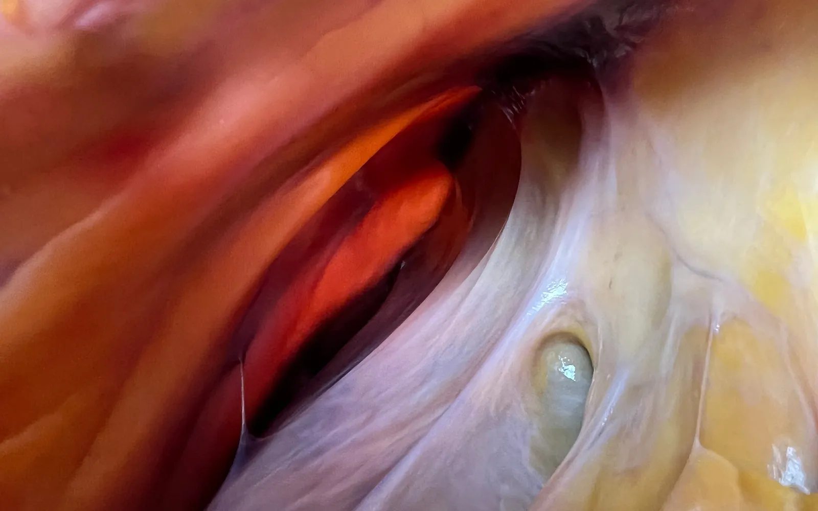
The thinner, inner layer of the serous pericardium is subdivided into the parietal layer, which is fused directly to the underside of the fibrous pericardium, and the visceral layer, called the epicardium, which hugs the surface of the heart muscle. This subdivision is what allows the heart to almost float as it beats. Lubricated by serous fluid, the slippery space between the two layers, called the pericardial cavity, minimizes friction as the heart smoothly pulses and twirls with every beat.
Unless you’ve gone looking specifically for images of the fibrous pericardium, you may never have seen it. When you see graphics or computer renderings of the heart beating, you typically only see the epicardium with the heart. You won’t see the fluid space or the fibrous pericardium. They will have been removed from the image because they completely obscure the view of the heart organ. This is a familiar story with fascial structures in the body. Fascia tends to cover and hide its more famous “cousins”—the muscles, bones, organs, vessels, and nerves.
Put your hand on your heart for a moment and picture how you imagine it lives in there.
Do you see the heart beating suspended in space? Or do you know about its container and how it’s situated within the pericardium, lying on its side?
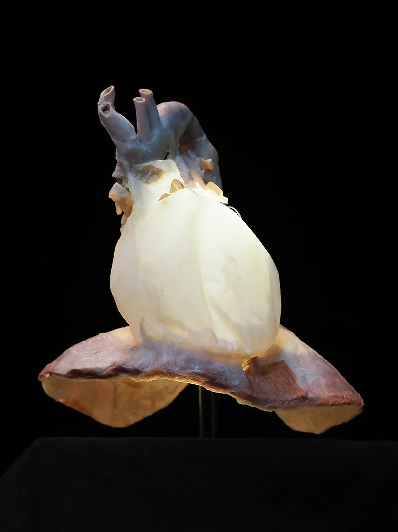
If all you can picture is the heart, you’re not alone. Students are often surprised at what we see when we open the chest cavity in our dissection labs. They look behind the sternum and ribs with confusion on their faces, expecting to see the heart and lungs. They can see the lungs, but the heart is nowhere to be found. Instead, in the center of the chest lies an unfamiliar, white, fibrous container that looks a bit like a vase, wider at the base, sitting on top of the muscular dome of the diaphragm. This white, vase-shaped column is the fibrous pericardium, a tough, relatively non-distensible connective tissue. The fibrous pericardium forms the container that holds the heart in place, and it has some pretty amazing connections.
The fibrous pericardium isn’t just a strong, protective sac loosely resting on the diaphragm; its base completely fuses with the central tendon of the diaphragm, forming a single, seamless fascial junction. This means every time you inhale deeply, your heart and pericardium go for a little ride, inferiorly, then superiorly. Down and up, all day long, thousands of times a day, millions of times a year; while it beats, your heart bounces along with the diaphragm’s movements.
Very few anatomy illustrations show this connection well, but it’s easily visible and a significant connection to note because it creates a stable continuity for how our breath affects our thoracic cavity three-dimensionally every time we breathe.
In addition to the inferior attachment to the diaphragm below, the pericardium is also connected superiorly to the fascia that surrounds the great vessels of the heart and the pretracheal fascia in the anterior neck. Posteriorly, it is connected to the esophagus, bronchi, and thoracic vertebral column (T5–T8). Anteriorly, the pericardium attaches firmly to the deep surface of the sternum. These strong connections in the chest add structural stability and cause the pericardium to slightly elongate with each breath.
Take another full breath. This time, as your lungs fill with air, visualize the dome of your diaphragm flattening and pulling downward with your inhale. The strong fascial covering of your heart is also pulled downward because of its connection with the diaphragm, while remaining stable through its other attachments superiorly, posteriorly, laterally, and anteriorly simultaneously. Take a few more breaths and notice how the diaphragm’s movement affects your torso three-dimensionally.
Understanding the heart-pericardium-diaphragm fascial connection helps us understand the three-dimensional body. Top to bottom, side to side, front to back, and from the center outward, we can access the pericardium-diaphragm link through breath, which allows us to reach deeper fascial connections than we ever could directly with our hands.
Superficial fascia may not get the same attention as deep fascia, but it plays an extremely important role in the organization of tissues and the structure of the body.
Understanding tendons—their shapes, lengths, and organization—improves an MT’s touch vocabulary and facilitates a more skilled touch.
While the neck is a bridge, a pathway, the position of the neck and head can also indicate a multitude of other things happening beneath the surface.
Understanding fibroblasts and the extracellular matrix changes how we think about the tissue we touch.