

Fascia: The Pericardium-Diaphragm Connection
Understanding how the pericardium relates to breathing can in turn help clients relax into their massage session.
The iliocostalis is part of the erector spinae (erect means “upright” and spinae means “spine”) group of muscles. The longissimus and spinalis are also part of this group. These muscles lie deep to the trapezius, latissimus dorsi, and rhomboids, and connect the sacrum, ilium, vertebral column, posterior ribs, and cranium. They provide broader stabilization and movement than the deeper transversospinalis group (rotatores and multifidi). Together, the erector spinae and transversospinalis groups maintain upright posture of the spine against gravity and position the vertebral column during movement.
The iliocostalis is the most lateral of the erector spinae muscles. Its fibers run from inferior to superior and medial to lateral, extending from the vertebral column out to the ribs like the branches of a tree. This branching strategy provides better leverage over long distances and protects the muscle fibers from shearing damage under stress. (This is demonstrated when a multitrunked, frequently branching tree more successfully resists wind damage. Greater frequency and directional diversity of branching offers more flexible and resilient trees, as well as better, safer motion control in the trunk.) When activated, the iliocostalis extends a flexed or laterally flexed trunk and controls flexion and lateral flexion against gravity.
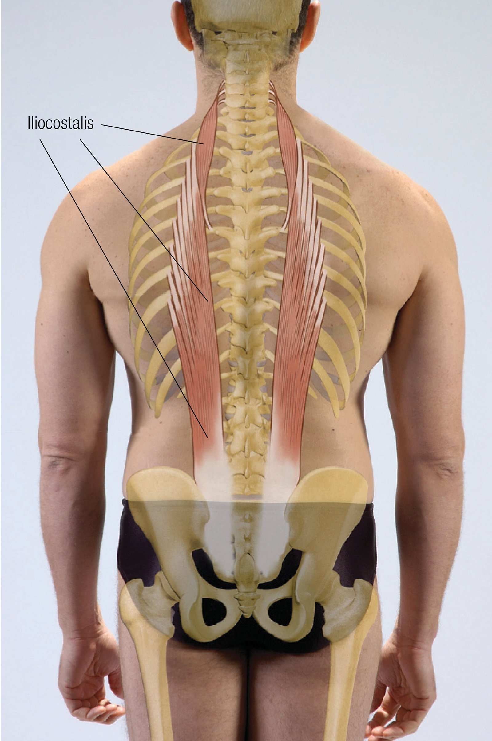
The spinalis and longissimus—the other erector spinae muscles—lie more medial and relatively vertical on the posterior trunk compared to the iliocostalis. This directionality limits their contribution to lateral flexion and rotation as more complex movements of the trunk are attempted. Additionally, the iliocostalis is the only erector spinae muscle that contributes to breathing, due to its attachments on the posterior ribs. Dysfunctional breathing patterns may contribute to pain and tension in the iliocostalis.
The overlapping layers of multidirectional muscles characteristic of the erector spinae and transversospinalis groups help stabilize and position the vertebral column through complex movements over great ranges of motion. The iliocostalis, with its long reach and branching strategy, provides the greatest amount of force production and control over the greatest diversity of movement. Balanced strength and mobility in the iliocostalis and the complex network of vertebral muscles is critical in maintaining healthy posture and safely and effectively navigating a wide variety of movements and activities. Reaching, bending, lifting, and even maintaining dynamic equilibrium during walking requires proper function of all of these muscles. Poor posture, improper body mechanics, functional or structural pelvic asymmetry, unilateral muscle tension or weakness, imbalanced utilization of the trunk muscles, and prolonged positioning or overuse of the upper extremities may cause pain and dysfunction in these muscles.
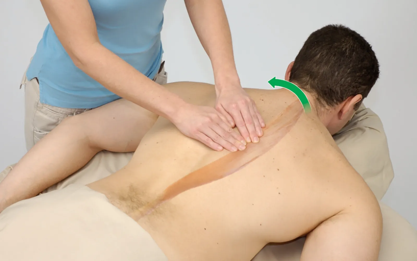
Positioning: client prone.
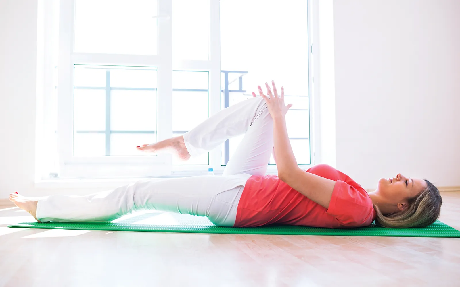
Editor’s note: The Client Homework element in Functional Anatomy is intended as a take-home resource for clients experiencing issues with the profiled muscle. The stretches identified in Functional Anatomy should not be performed within massage sessions or progressed by massage therapists, in order to comply with state laws and maintain scope of practice.
Understanding how the pericardium relates to breathing can in turn help clients relax into their massage session.
Superficial fascia may not get the same attention as deep fascia, but it plays an extremely important role in the organization of tissues and the structure of the body.
Understanding tendons—their shapes, lengths, and organization—improves an MT’s touch vocabulary and facilitates a more skilled touch.
While the neck is a bridge, a pathway, the position of the neck and head can also indicate a multitude of other things happening beneath the surface.