

Fascia: The Pericardium-Diaphragm Connection
Understanding how the pericardium relates to breathing can in turn help clients relax into their massage session.
In the last edition of "Functional Anatomy" (March/April 2016), we expanded our focus beyond muscles and muscle groups to include other structures affected by bodywork. We began with an examination of the muscle spindle and continue here with the Golgi tendon organs.
Like the muscle spindles we explored last time, Golgi tendon organs are proprioceptors and contribute to an overall awareness of body position. Various sensory structures—in the joints, muscles, and tendons, including Golgi tendon organs—communicate positional information to the central nervous system in an effort to maintain balanced posture, create smooth and efficient movement, and prevent injury.
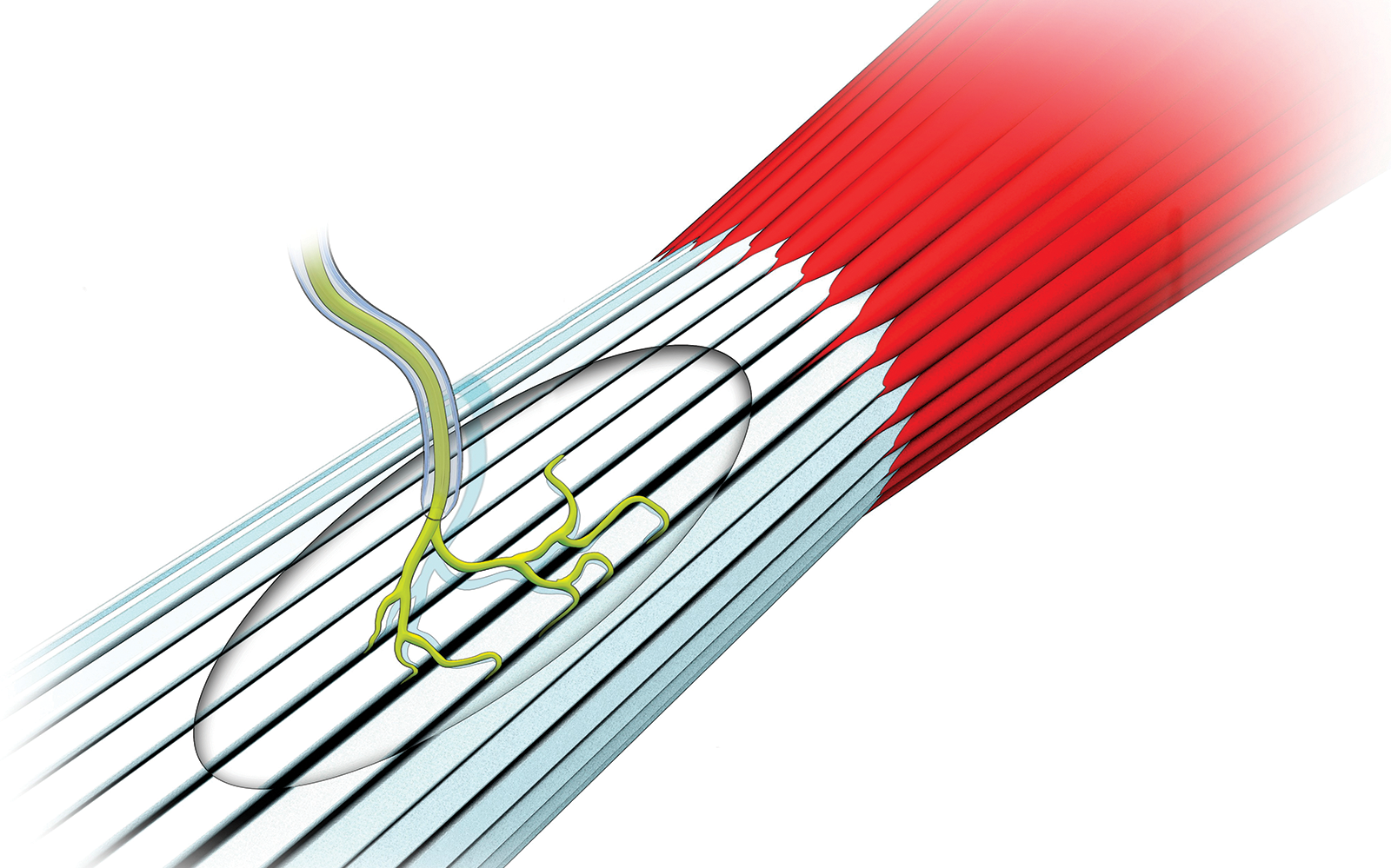
Golgi tendon organs are sensory structures woven into tissues at the musculotendinous junction near the origins and insertions of skeletal muscles. This location is slightly different than that of muscle spindles, which are located within the muscle belly. Each Golgi tendon organ is made of collagen intrafusal fibers that run parallel to the tendon at one end and extend into the muscle belly at the other. A dense capsule of extrafusal fibers, which are tightly packed strands of collagen, encloses the Golgi tendon organ. A single afferent nerve fiber within the capsule branches out and spirals its terminal ends around individual convoluted intrafusal fibers and detects changes in tissue.
Where the muscle spindles monitor the rate of length change when muscles stretch, the Golgi tendon organs monitor changes in muscle tension. Muscle tension is created when muscles lengthen and stretch, and also when they are activated to create isometric or isotonic muscle contractions. As muscle tension increases, the sensory terminals of the afferent nerve are compressed. This compression initiates communication with the central nervous system regarding the amount of tension generated in the muscle.
If safe amounts of muscle tension are generated, the Golgi tendon organs will initiate an action called reciprocal inhibition. Reciprocal inhibition describes the relaxation of one muscle while the opposite is activated. When you lift an object of reasonable weight by gripping it and bending or flexing your elbow, the muscles that extend your fingers and elbow relax, allowing the motion to happen without resistance. This coordinated muscle activation and relaxation allows the body to move efficiently and not fight against itself. Appropriate give-and-take must occur between opposing muscle groups in order for smooth, coordinated movement to take place.
Golgi tendon organs respond differently if muscle tension is perceived to be excessive or dangerous. In these situations, they initiate the inverse myotatic reflex. When muscles generate excessive tension, the Golgi tendon organs will inhibit further muscle activation and force the muscles to relax. It also prompts the opposite muscle group to contract and shorten. Both actions decrease tension on the affected muscle. We see this response in cliffhanger movies when someone is hanging on for dear life and then his fingers just let go. This letting go is a function of the Golgi tendon organs inhibiting muscle activation to the overtense flexors, while simultaneously activating the opposing extensors, thus opening the hand.
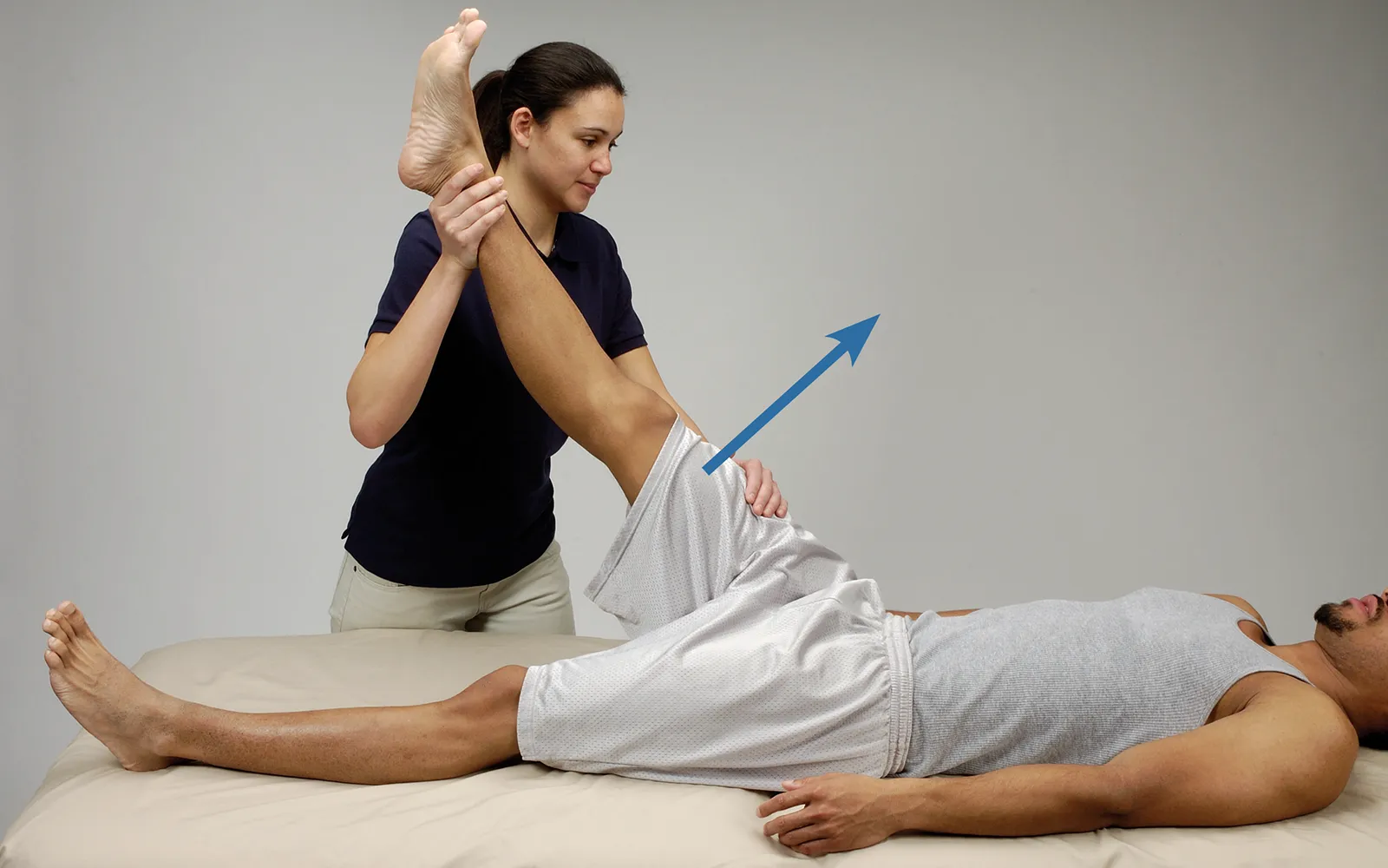
One option is to utilize reciprocal inhibition to suppress activation of a target muscle. This is particularly useful to combat muscle cramps and reduce hypertonicity and guarding. The method uses isometric contraction of an opposing muscle or muscle group to prompt relaxation of the target muscle.
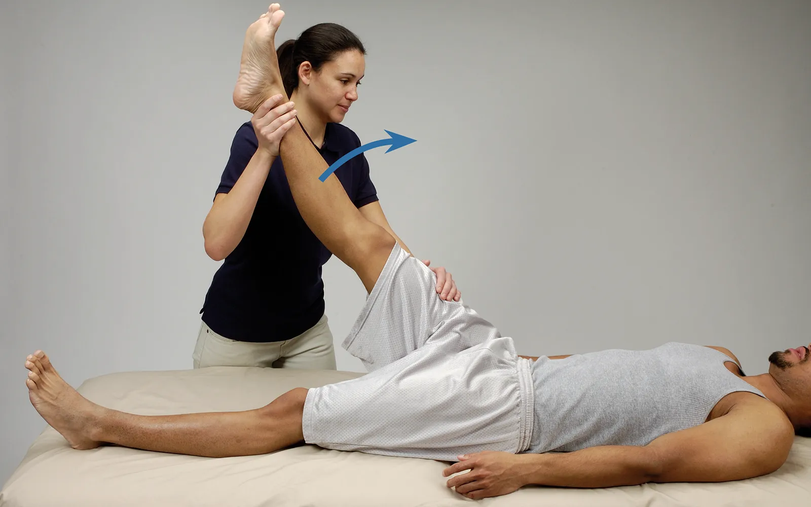
Positioning: client supine, support leg with knee straight and hip passively flexed.
Understanding how the pericardium relates to breathing can in turn help clients relax into their massage session.
Superficial fascia may not get the same attention as deep fascia, but it plays an extremely important role in the organization of tissues and the structure of the body.
Understanding tendons—their shapes, lengths, and organization—improves an MT’s touch vocabulary and facilitates a more skilled touch.
While the neck is a bridge, a pathway, the position of the neck and head can also indicate a multitude of other things happening beneath the surface.