

When Superficial Fascia Goes Deep
Superficial fascia may not get the same attention as deep fascia, but it plays an extremely important role in the organization of tissues and the structure of the body.
Let's continue to examine structures that distribute forces and absorb impact in the human body. In previous issues, we explored the architecture and function of the pelvic girdle ("Pelvic Girdle Suspension System," November/December 2017, page 41) and the foot ("Force Distribution in the Foot," January/February 2018). We continue here with the structure and function of the intervertebral discs.
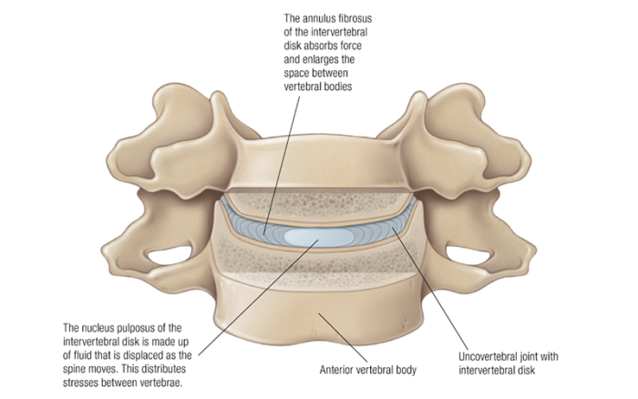
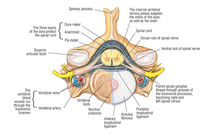
Intervertebral discs are the rounded layers of connective tissue that lie between each vertebra in the human vertebral column. Each disc is composed of three distinct segments: a fluid center called the nucleus pulposus; a tough, fibrous outer layer called the annulus fibrosus; and thin layers of cartilaginous end plates at the top and bottom. Each segment serves a particular role in fulfilling the overall function of maintaining space between adjacent vertebrae, while simultaneously distributing compressive forces through the vertebral column.
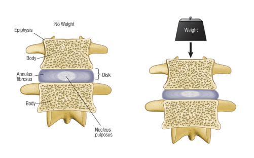
The nucleus pulposus is a gelatinous segment that contains high levels of proteoglycans and water with lesser amounts of collagen. The water content is variable and chemically controlled by living cells. These cells produce proteoglycans, chemicals that attract and hold water. Healthy intervertebral discs are able to adjust hydrostatic pressure and water content in response to varying amounts of mechanical compression or loading, thus evenly and optimally distributing loads to surrounding structures. It is the high water content that gives the disc flexibility and "height," ensuring adequate space for nutrient diffusion within the disc and preventing compression or impingement of associated soft tissues like nerve roots located between vertebrae.
The annulus fibrosus is a tough, dense layer of fibrocartilage that contains much higher amounts of collagen than the nucleus pulposus. A complex network of collagen and other supportive fibers provides resistance to tensile forces, elasticity, and architectural "firmness" to the disc. Proper orientation of the fibers protects and properly distributes compressive forces placed on the nucleus pulposus. Though less prevalent than in the nucleus pulposus, proteoglycan production and associated water content is critical to the health and proper function of the annulus fibrosus.
Encouraging movement at individual spinal segments optimizes nutrient flow and minimizes excessive loading on a given segment.
The cartilaginous end plates are thin layers of hyaline cartilage at the superior and inferior margins of the intervertebral disc. They provide connection between the intervertebral disc and the associated vertebral bodies. These regions contain the least amount of water and greatest amount of resilient collagen fibers, providing a relatively stable connection while maintaining optimal nutrient diffusion between the disc and adjacent bony tissues.
Intervertebral discs are avascular (lack blood supply) and, therefore, the living cells within rely on diffusion of water, ions, and small molecules for nutrient delivery. Like all connective tissues, the intervertebral discs contain living cells within an extracellular matrix. These cells need energy and raw materials in order to assess, synthesize, and break down the extracellular matrix, which is how they continuously repair and adjust the intervertebral disc architecture for optimal function.
Excessive loading—either in scale or duration—disrupts the flow of nutrients within the intervertebral disc, leading to cell death and a cascade of changes within the tissue. Decreased proteoglycan production decreases water content within the tissue. The tissue becomes less resistant to compression, showing greater rates of fluid loss with loading. Disc height reduces as the tissue becomes more fibrotic and the percentage of collagen fibers increases, causing ineffective load distribution and compromise of surrounding structures.
Since muscle tension places the greatest amount of loading on the spine, it is essential to evenly and optimally direct that loading. Focus should be placed on correcting postural deviations that asymmetrically load the spine. Also, encouraging movement at individual spinal segments optimizes nutrient flow and minimizes excessive loading on a given segment. Balanced tension between antagonistic muscle groups and reduced hypertonicity in both the intervertebral and paravertebral muscle groups reduces loading on the intervertebral discs and supports nutrient flow.
Superficial fascia may not get the same attention as deep fascia, but it plays an extremely important role in the organization of tissues and the structure of the body.
Understanding tendons—their shapes, lengths, and organization—improves an MT’s touch vocabulary and facilitates a more skilled touch.
While the neck is a bridge, a pathway, the position of the neck and head can also indicate a multitude of other things happening beneath the surface.
Understanding fibroblasts and the extracellular matrix changes how we think about the tissue we touch.