

Fascia: The Pericardium-Diaphragm Connection
Understanding how the pericardium relates to breathing can in turn help clients relax into their massage session.
The platysma is the most superficial muscle of the anterior neck, and extends from the mandible and fascia of the face. Its origin is not bony; rather, it is attached to the fascia of the chest and the anterior shoulder girdle. This muscle is one of several mimetic muscles, or muscles responsible for facial expression. Each of these muscles is controlled by the facial nerve, with the platysma specifically controlled by the deep cervical portion.
The platysma is a flat, continuous sheet of muscle (generally termed a panniculus muscle). Panniculus muscles are found in many animals, and function to move areas of skin, a movement useful in shaking off flies, raising hair, and shivering. In humans, the platysma is a vestigial muscle made redundant by the broad range of the upper extremities, which allows the hand to reach nearly all parts of the body.
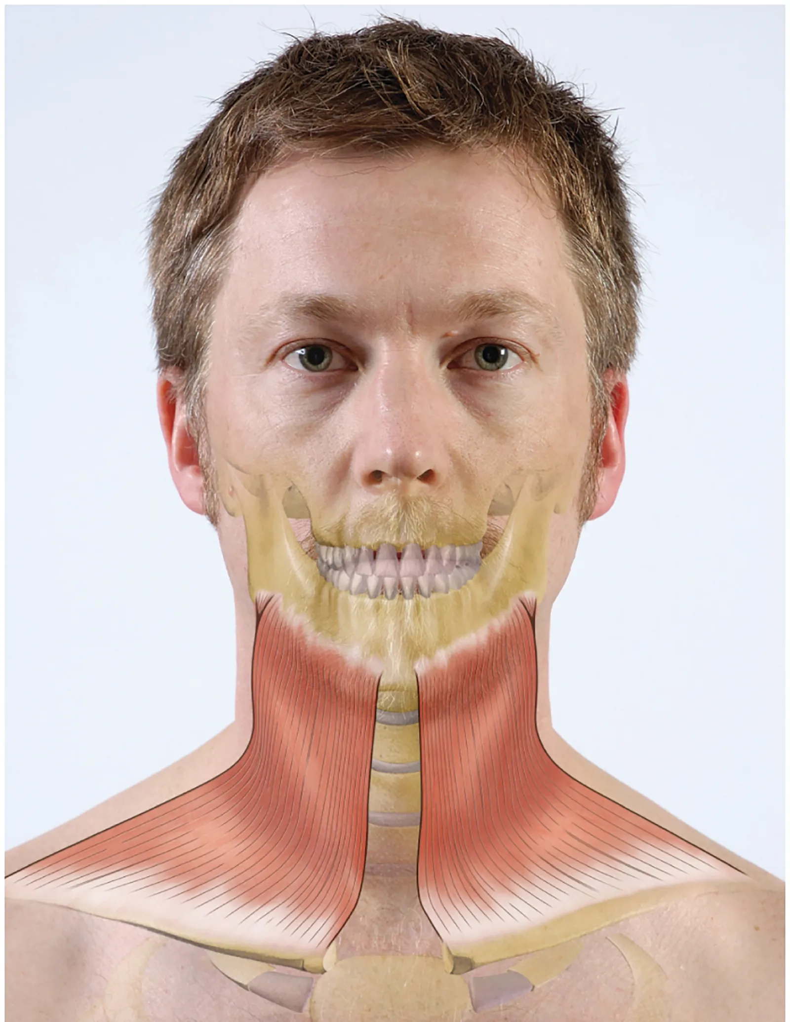
While the platysma is less functional in flicking flies, it is very active in creating specific human facial expressions. This muscle draws the lower lip downward and laterally while creating ridges or wrinkles in the skin of the neck and chest. When combined with different movements of the forehead, eyes, and nose, this action characterizes the facial expressions of stress or anger.
Prolonged periods of deep concentration or stress may create tension and pain in the platysma. This tension may be exacerbated by poor tissue mobility around the pectoralis major and anterior deltoid, as well as postural deviations like forward head posture or kyphosis. Improving posture in the head, neck, and shoulder girdle, as well as circulation and tissue mobility in the chest and anterior shoulder, may alleviate pain and tension in the platysma.
Clients who must emphasize or exaggerate facial expressions, like actors, performers, sign language interpreters, and teachers, may experience excessive tension in the platysma, as well as the other mimetic muscles. Include superficial friction techniques and myofascial release of these muscles for these clients in particular, or as part of treatment for neck issues, temporomandibular joint dysfunction, headaches, or as a nice finish for relaxation massage.
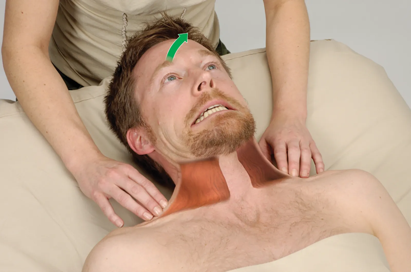
Positioning: client supine.
Editor’s note: The Client Homework element in Functional Anatomy is intended as a take-home resource for clients experiencing issues with the profiled muscle. The stretches identified in Functional Anatomy should not be performed within massage sessions or progressed by massage therapists, in order to comply with state laws and maintain scope of practice.
Understanding how the pericardium relates to breathing can in turn help clients relax into their massage session.
Superficial fascia may not get the same attention as deep fascia, but it plays an extremely important role in the organization of tissues and the structure of the body.
Understanding tendons—their shapes, lengths, and organization—improves an MT’s touch vocabulary and facilitates a more skilled touch.
While the neck is a bridge, a pathway, the position of the neck and head can also indicate a multitude of other things happening beneath the surface.