

Going for Gold
A gold medal-winning massage may not be what you think. It’s not about fancy tools, flashy techniques, or pedigree. It’s about centering the bodywork on the client experience.
Ida Rolf once said, “Where you think the pain is, the problem is not.” This holds true in the worlds of osteopathy, physical therapy, massage, and bodywork. The following case study, taken from my own physical therapy clinic at the University of Oxford, illustrates Rolf’s statement well. The patient presented with pain in her left shoulder, but after careful examination, evaluation, and detective work, it was determined that the cause of the problem originated in an area one might not have originally considered.
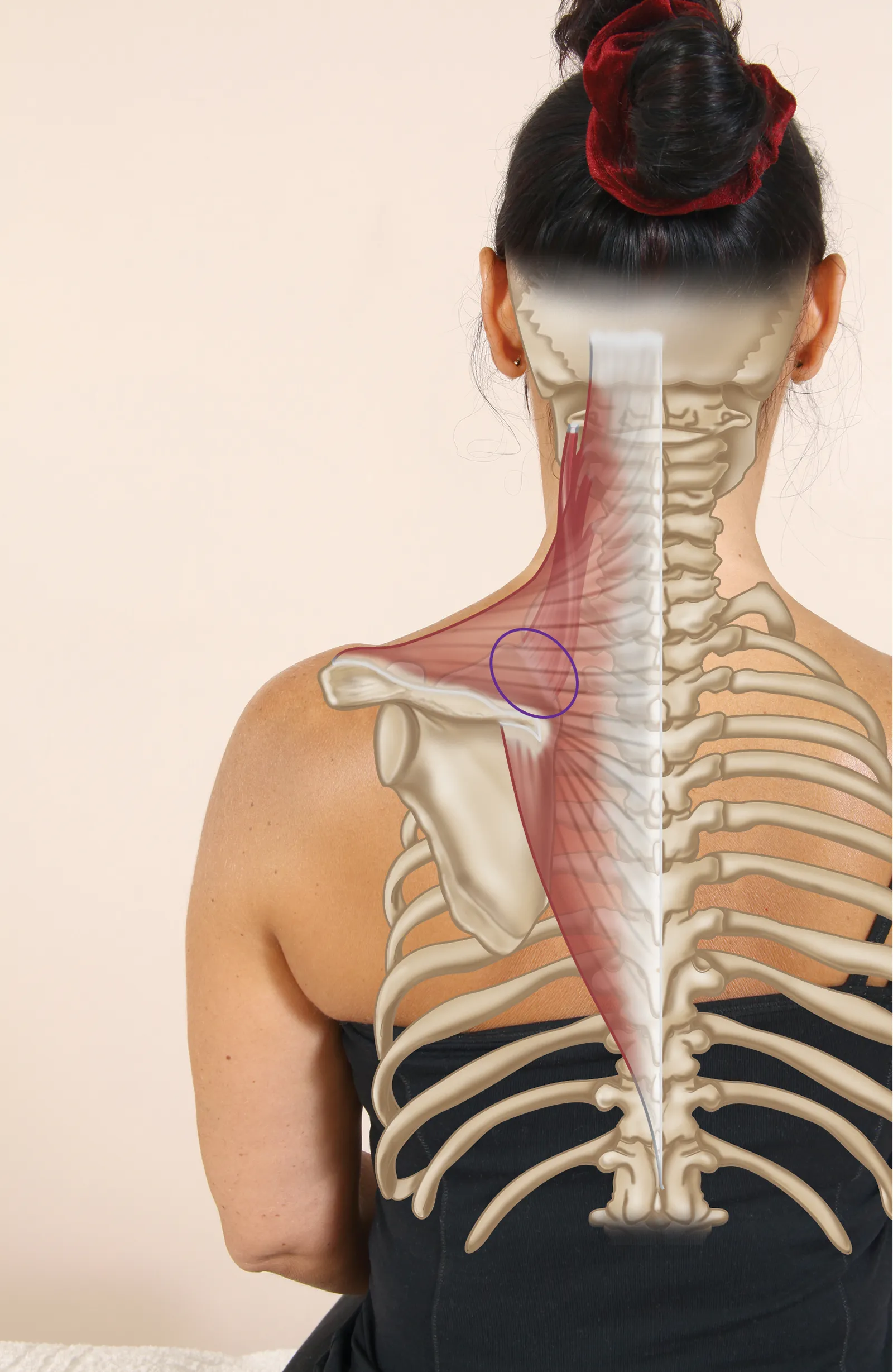
The patient in question was a 34-year-old female physical trainer for the Royal Air Force. She presented to the clinic with pain near the superior aspect of her left scapula (Image 1). The pain would present at the 4-mile mark of a run, forcing her to stop because it was so intense. The discomfort would then subside, but quickly return if she attempted to start running again. Running was the only activity that caused the pain. Her complaint had been ongoing for eight months, had worsened over the past three, and was starting to affect her work. There was no previous history or related trauma to trigger the complaint.
After seeing different practitioners, who all focused their treatment on the upper trapezius, she visited an osteopath who treated her cervical spine and rib area. The treatments she received included the application of soft-tissue techniques to the affected area—namely the trapezius, levator scapulae, sternocleidomastoid (SCM), scalenes, etc. The osteopath also used manipulative techniques on the facet joints of her cervical spine: C4–5 and C5–6. Muscle energy techniques and trigger-point releases were used in a localized area, which offered relief at the time, but made no difference when she attempted to run more than 4 miles. She had not undergone any scans (e.g., MRI or X-ray).
When the patient came for a consultation at my clinic, the physical therapist working the case asked about the woman’s pain. She reported that the potential tissues responsible for the pain in her superior scapula were the upper trapezius, levator scapulae, scalenes, thoracic rib, and cervical rib.
Once a subjective history was conducted, an objective assessment began, including active range of motion, passive range of motion, resisted range of motion, and palpation tests to determine the condition of the affected tissues.
When considering this patient’s case, there were a variety of potential causes of her pain ranging from dysfunction of the glenohumeral or the acromioclavicular joint to an elevated first rib.
When I see a new patient for the first time, no matter what the presenting pain is, I normally assess the pelvis for position and movement, as I consider this area of the body in particular to be the foundation for everything that connects to it. I often find that when I correct a dysfunctional pelvis, the patient’s presenting symptoms tend to settle down. However, when I assessed this particular patient, I found her pelvis was level and moving correctly. I then went on to test the firing patterns of the gluteus maximus, which I often do with patients and athletes who participate in regular athletic activities.
However, I only test the firing pattern sequence once I feel that the pelvis is in its correct position; the logic here is that you often get a positive result of the muscle misfiring when the pelvis is slightly out of position.
With the patient in question, I found a bilateral weakness/misfiring of the gluteus maximus, but the firing on the right side seemed a bit slower. As I had not found any dysfunction in the pelvis, I pursued this line of approach a little further.
Before we continue, I would like to pose a few questions for you to think about:
To answer these questions, we need to put on our detective hats and look at the functional anatomy of the gluteus maximus, as well as the relationship of the gluteus maximus to other anatomical structures.
The gluteus maximus operates mainly as a powerful hip extensor and a lateral rotator, but it also plays a part in stabilizing the sacroiliac joint (SIJ) by helping it to “force close” while going through the gait cycle.
Some of the gluteus maximus muscular fibers attach to the sacrotuberous ligament, which runs from the sacrum to the ischial tuberosity. This ligament has been termed the key ligament in helping to stabilize the SIJ. To gain a better understanding of this action, we first need to consider two concepts—form closure and force closure—that are both associated with stability of the SIJ.
The shape of the sacrum—along with its ridges and grooves and the fact that it is wedged between the ilia—helps to bring natural stability to the SIJ. This is known as form closure. If the articular surfaces of the sacrum and the ilia fit together with perfect form closure, mobility would be practically nonexistent. However, form closure of the SIJ is not perfect and movement is possible, which means stabilization during loading is required. This is achieved by increasing compression across the joint at the moment of loading; the surrounding ligaments, muscles, and fascia are responsible for this. The mechanism of compression of the SIJ by these additional forces is called force closure.
When the body is working efficiently, the forces between the innominates and the sacrum are adequately controlled, and loads can be transferred between the trunk, pelvis, and legs. So, how do we link this to the patient’s complaint? The posterior oblique sling directly links the right gluteus maximus to the left latissimus dorsi via the thoracolumbar fascia. The latissimus dorsi has its insertion on the inner part of the humerus, and one of the functions of this muscle is to keep the scapula against the thoracic rib cage and aid in depression of the scapula.
So what do we know? We know that the right side of the patient’s gluteus maximus is slightly slower in terms of its firing pattern, and that this muscle plays a role in the force closure process of the SIJ. This tells us that if the gluteus maximus cannot perform this function of stabilizing the SIJ, then something else will assist in stabilizing the joint. The left latissimus dorsi is the synergist that helps stabilize the right gluteus maximus and, more importantly, the SIJ. When the patient runs, every time her right leg contacts the ground and goes through the gait cycle, the left latissimus dorsi is overcontracting. This causes the left scapula to depress, and the muscles that resist the downward depressive pull will be the upper trapezius and the levator scapulae. Subsequently, these muscles start to fatigue. For the patient in question, this fatigue occurs at approximately 4 miles, at which point she feels pain in her left superior scapula.
You might think the easy way to treat a weakness in the gluteus maximus is to encourage your client to do strength-based exercises. However, in practice, this is not always the correct solution, as sometimes the tighter antagonistic muscle is responsible for the apparent weakness. The muscle in this case is the iliopsoas (hip flexor), and the shortening of this can result in a weakness inhibition of the gluteus maximus. My answer to this puzzle was to stretch the patient’s right iliopsoas muscle to see if it promoted the firing activation of the gluteus maximus, while at the same time introducing strength exercises for the gluteus maximus.
I advised the patient to abstain from running and to get her partner to assist in lengthening the iliopsoas, rectus femoris, and adductors twice a day (see the iliopsoas technique described below). Strength exercises were also advised twice daily until the follow-up treatment. I reassessed her 10 days later and found normal firing of the gluteus maximus on the hip extension firing pattern test, and a reduction in the tightness of the associated iliopsoas, rectus femoris, and adductors. Because of these positive results, I advised her to run as far as felt comfortable. I was not sure if my treatment was going to correct the problem, but she reported she had no pain during, or after, a 6-mile run. The patient is still pain-free and continues to regularly use gluteus maximus strengthening exercises and lengthening techniques for the tight muscles.
This case study demonstrates that often the underlying cause of a condition or problem may not be local to where the symptoms/pain present, which means that all avenues need to be fully considered. Remember, physical therapy, like massage therapy, is much like a jigsaw puzzle—if you stick with it, the picture will eventually become a lot clearer.
Since I am discussing how to maximize the glutes through lengthening the tight antagonist muscles, I need to explain the role of these techniques so you have a better understanding of when and why to employ this type of treatment with your clients.
Muscle energy techniques (METs) are employed by massage therapists, physical therapists, osteopaths, sports therapists, and others to lengthen postural muscles that are prone to shortening. The primary force in an MET comes from the contraction of the client’s soft tissues, which is then utilized to assist and correct the presenting musculoskeletal dysfunction.
When teaching the concept of METs to my students, one of the benefits I emphasize is their use in normalizing joint range, rather than improving flexibility. This is not stretching in the strictest sense—even though overall flexibility will improve, it is only to the point of achieving what is considered to be a normal joint range. Depending on the context and the type of MET employed, the objectives of this treatment can include restoring normal tone in hypertonic muscles, strengthening weak muscles, preparing muscles for subsequent stretching, and increasing joint mobility.
When applying METs, it’s important to note that the point of bind, or restriction barrier, occurs when resistance is first felt by the palpating hand/fingers of the therapist. Through experience and continual practice, the therapist will be able to palpate a resistance of the soft tissues as the affected area is gently taken into the position of bind. This position of bind is not the position of stretch—it is the position just before the point of stretch. The therapist should be able to feel the difference and not wait for the client to say when they feel a stretch has occurred.
When working with the glutes, it’s important to remember which muscles are antagonistic to this muscle group. The gluteus maximus, in particular, is a powerful hip extensor, so its antagonist has to be the hip flexors—the main muscles responsible for hip flexion, specifically the iliopsoas (psoas major and iliacus) and rectus femoris. The gluteus medius is a powerful hip abductor, so its antagonist has to be the adductors. One way of encouraging a correct hip extension firing pattern is to identify and correct a hip flexor length issue: if the flexors are tested as short, an MET or a myofascial release technique can be utilized to normalize the resting length of these shortened structures. This process of lengthening the shortened structures can be applied for a period of approximately two weeks; if the specific firing pattern has not improved after this time, strengthening protocols for the gluteus maximus can then be incorporated into the treatment plan.
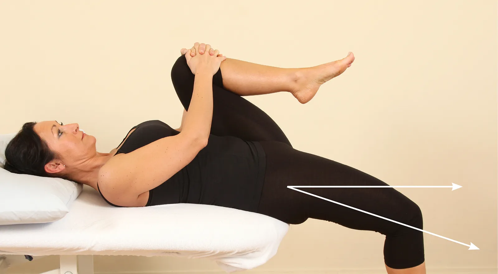
To test the right hip, the client is asked to lie back on the edge of the table while holding onto her left knee. As she rolls backward, the client pulls her left knee as far as she can toward her chest (Image 2). The full flexion of the hip encourages full posterior rotation of the innominate bone and helps to flatten the lordosis. From this position, the therapist looks at where the client’s right knee lies relative to the right hip. The position of the knee should be just below the level of the hip; Image 2 demonstrates a normal length of the right iliopsoas.
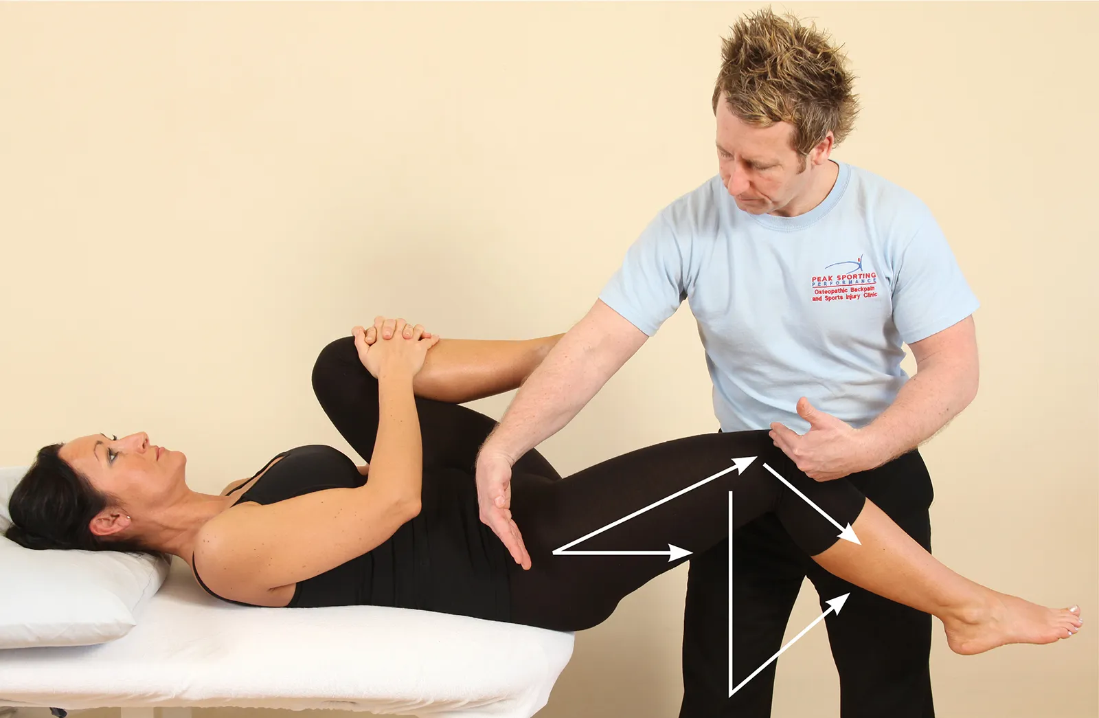
In Image 3, the therapist is demonstrating with his arms the position of the right hip compared with the right knee. You can see that the hip is held in a flexed position, which confirms the tightness of the right iliopsoas in this case.
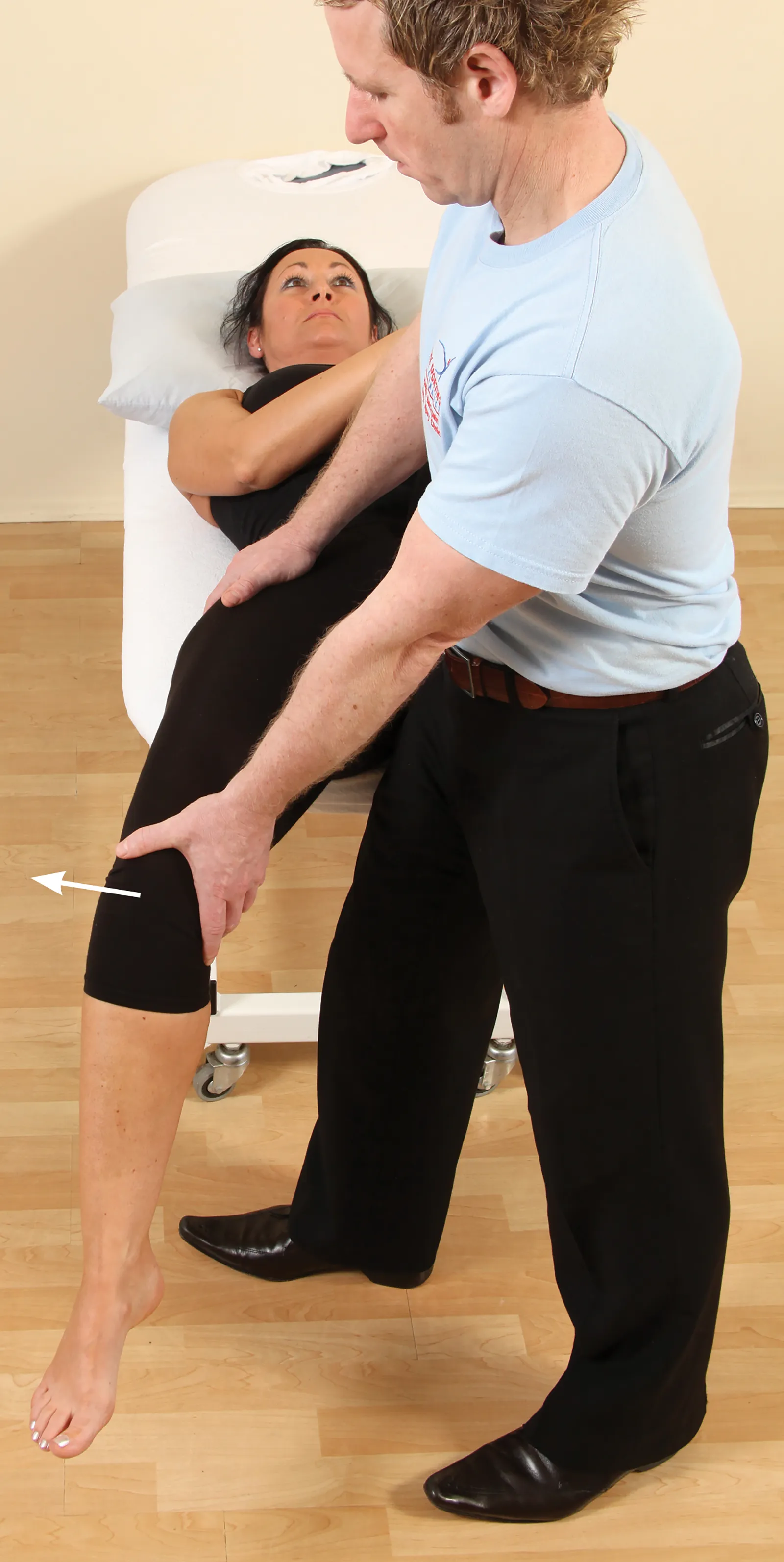
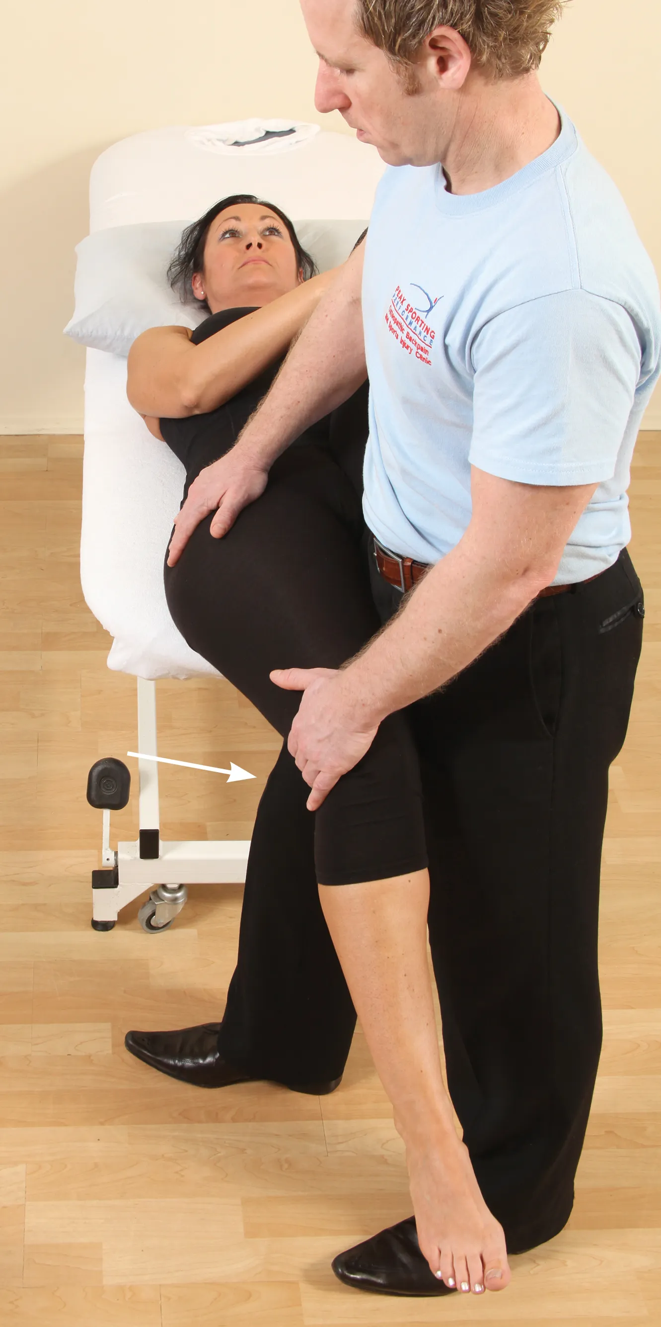
With the client in the Modified Thomas Test position, the therapist can apply an abduction of the hip (Image 4), and an adduction of the hip (Image 5). A range of movement (ROM) of 10–15 degrees for each of these is commonly accepted to be normal. If the hip is restricted in abduction (i.e., a bind occurs at an angle of less than 10–15 degrees), the muscles of the adductor group are held in a shortened position; if the adduction movement is restricted, the iliotibial band (ITB) and tensor fasciae latae (TFL) are held in a shortened position.
To treat the right side, the client adopts the same position as in the Modified Thomas Test. The client’s left foot is placed into the therapist’s right side and pressure is applied by the therapist to induce full flexion of the client’s left hip. Stabilizing the client’s right hip with his right hand, the therapist also puts his left hand just above the client’s right knee. The client is asked to flex her right hip against the therapist’s resistance for 10 seconds, as shown in Image 6.
Following the isometric contraction, and on the relaxation phase, the therapist slowly applies a downward pressure. This will cause the hip to passively go into extension and will induce a lengthening of the right iliopsoas, as shown in Image 7. Gravity will also play a part in this technique, by assisting in the lengthening of the iliopsoas.
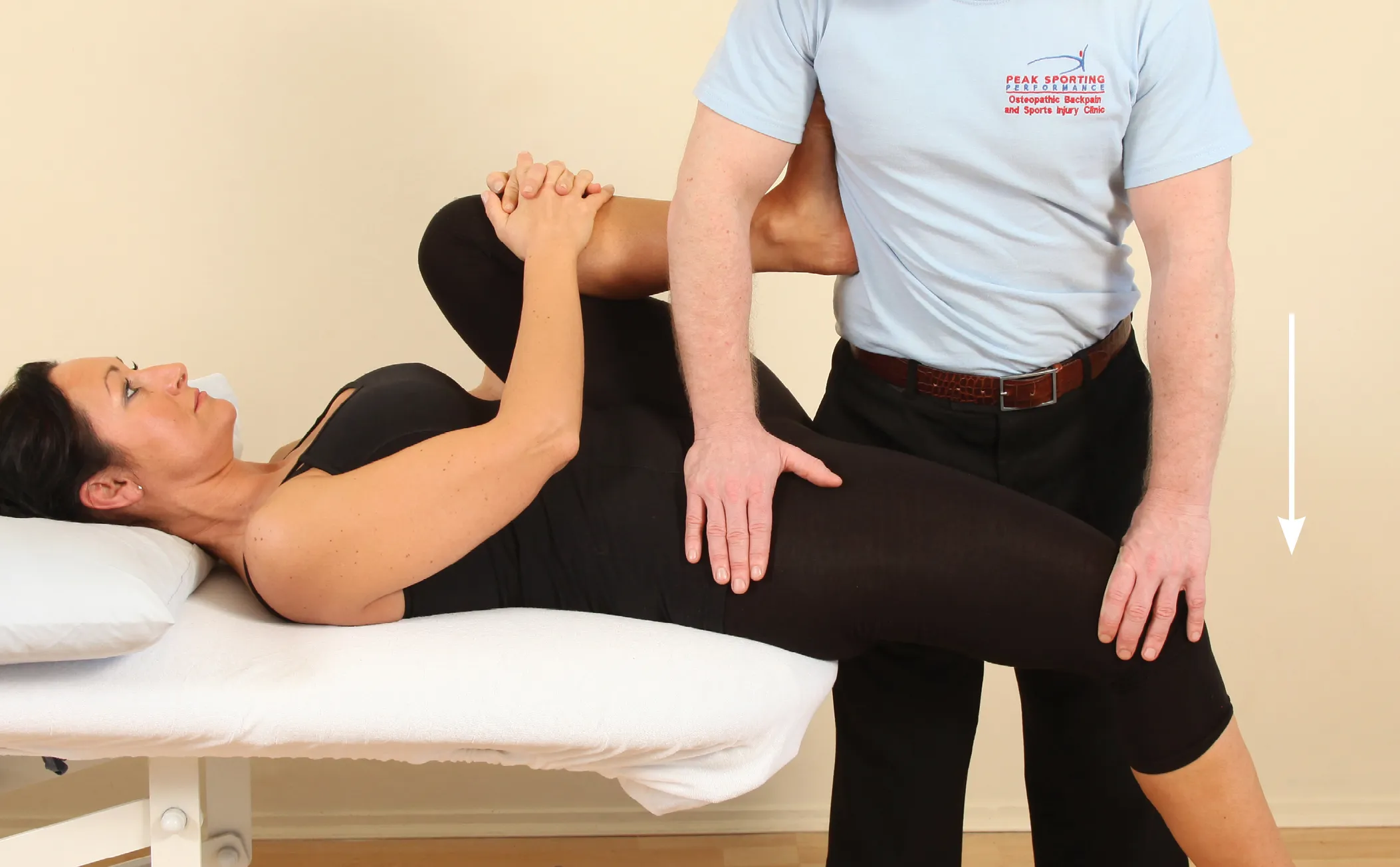
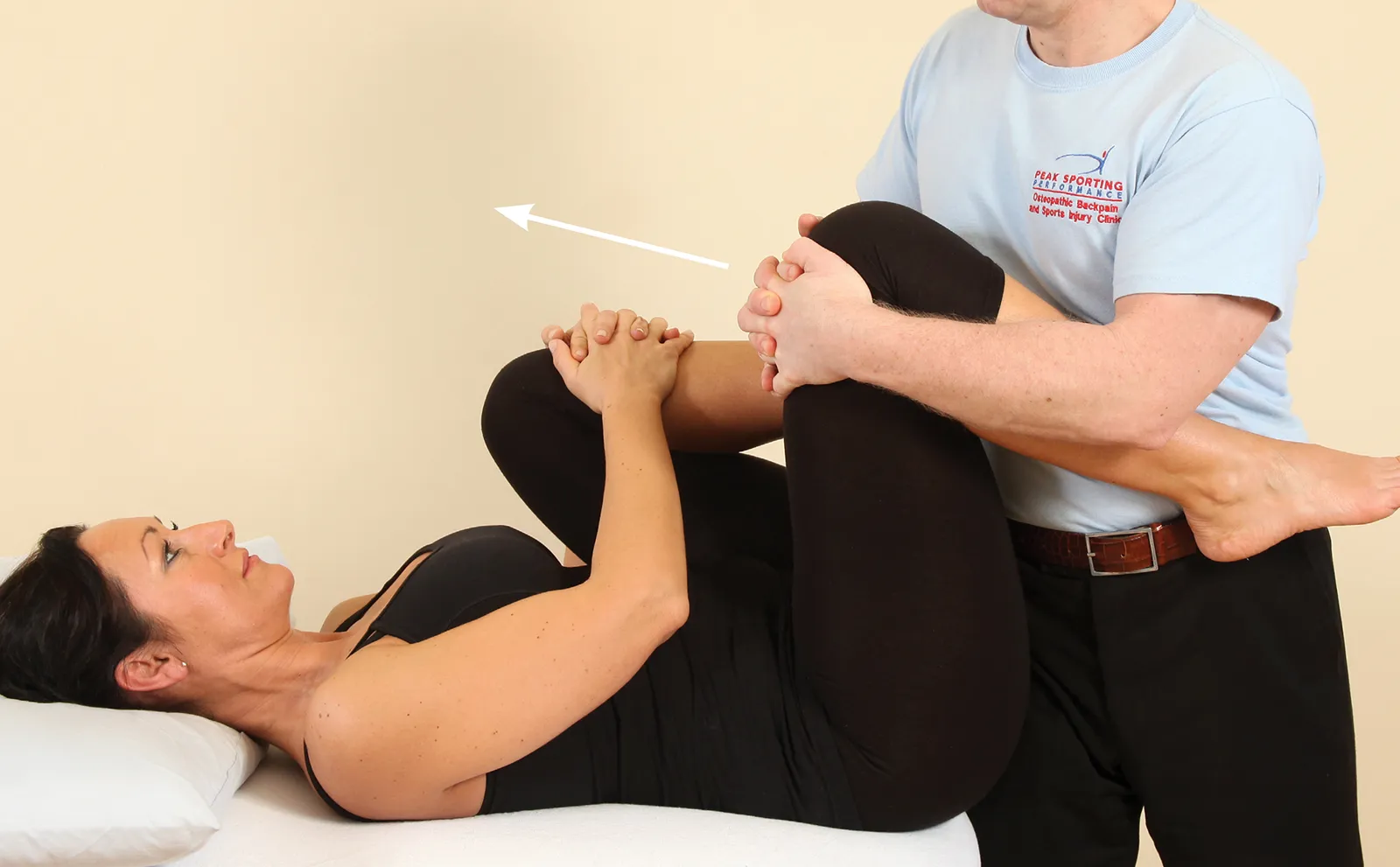
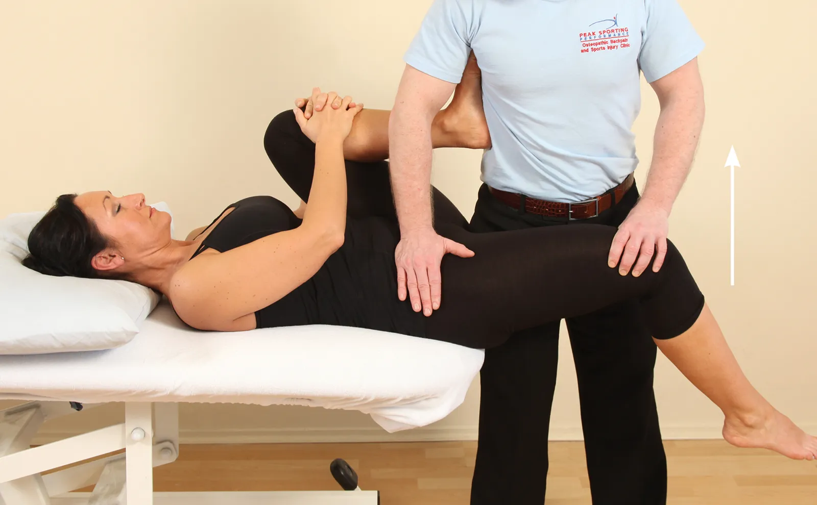
Alternatively, it is possible to contract the iliopsoas from the flexed position, as shown in Image 8. This is normally used if the original method of activating the iliopsoas causes discomfort to the client. Allowing the hip to be in a more flexed position will slacken the iliopsoas, which will assist in its contraction and help reduce the discomfort.
The client is asked to flex her right hip against a resistance applied by the therapist’s left hand (Image 8). After a 10-second contraction, and on the relaxation phase, the therapist lengthens the iliopsoas by returning the hip to the extended position shown in Image 7.
This type of technique is excellent for relaxing and releasing tone in tight, shortened soft tissues.
A gold medal-winning massage may not be what you think. It’s not about fancy tools, flashy techniques, or pedigree. It’s about centering the bodywork on the client experience.
Energy work can be a divisive topic in the massage field, however, there are valuable potential benefits and insights to be gained through exploring and integrating energetic modalities in therapeutic practice.
Time perception is shaped by interoception and body state, which means hands-on work can directly influence how clients experience duration.
By recognizing the knee as a transmission point within the kinetic chain rather than a standalone joint, therapists can address the upstream and downstream restrictions that can cause knee stress and pain.