

Fascia: The Pericardium-Diaphragm Connection
Understanding how the pericardium relates to breathing can in turn help clients relax into their massage session.
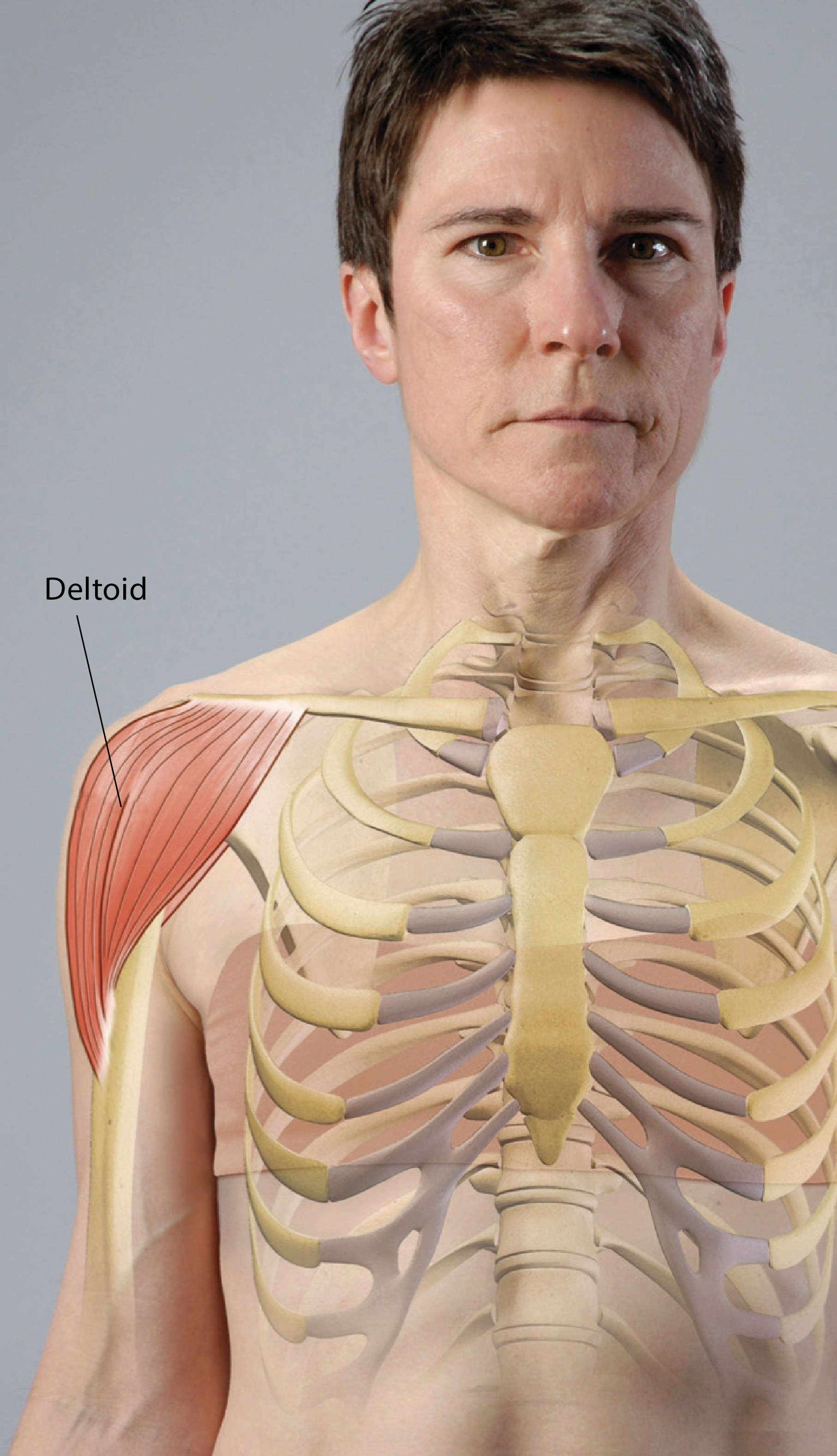
The deltoid muscle is a large, superficial structure that covers the lateral shoulder. The muscle originates at the lateral one-third of the clavicle anteriorly, the acromion process laterally, and the spine of the scapula posteriorly. It begins where the trapezius muscle ends, but with clear delineation provided by the bony landmarks. The thick fibers of the deltoid converge to a single point halfway down the lateral side of the humerus and insert on the deltoid tuberosity. The muscle is named for the triangular shape it forms on the shoulder.
The deltoid is a prime mover for nearly all movements of the shoulder. Its multipennate fiber arrangement, large cross-sectional area, and broad attachment points create excellent leverage on the glenohumeral joint. The deltoid also plays an important role in stabilizing the shoulder by wrapping itself around the glenohumeral joint and holding it together. When all fibers of the deltoid work together, it is a powerful abductor. The supraspinatus stabilizes the head of the humerus as the deltoid abducts the shoulder and prevents impingement of the humeral head on the acromion process. This tandem action provides the smooth and powerful movement of the shoulder necessary for lifting and reaching both above and below shoulder height.
The anterior fibers of the deltoid work with the pectoralis major to flex, horizontally adduct, and internally rotate the humerus. This is a powerful combination, and these muscles are utilized in pushing, reaching, and initiating throwing movements. As a result of this association with the pectoralis major, as well as the fact that most activities of daily living utilize movements in front of the body, the anterior fibers of the deltoid are often overdeveloped and the posterior fibers are typically underdeveloped. The anterior deltoid frequently adheres to the pectoralis major where the muscles overlap at the anterior shoulder. This pattern contributes to mechanical dysfunction, soft-tissue impingement, and pain in the glenohumeral joint and associated structures.
The posterior fibers of the deltoid are antagonists to the anterior fibers in all motions except abduction. These fibers horizontally abduct and externally rotate the shoulder. They also work with the latissimus dorsi and teres major to extend the shoulder, with the pectoralis major contributing to this motion from overhead, flexed positions. Functionally, these movements are critical for pulling and overhand throwing motions. Effective and efficient movement of the glenohumeral joint depends on proper coordination and development of all fibers of the deltoid and the muscles of the rotator cuff.
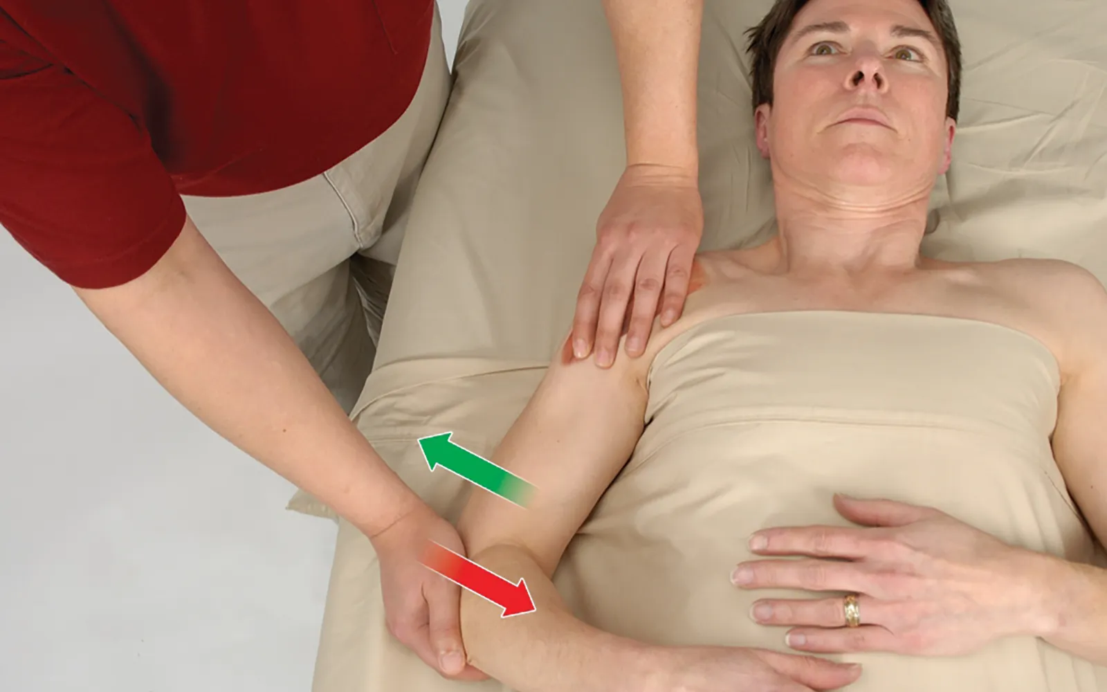
Positioning: client supine with arm at side.
Editor’s note: The Client Homework element in Functional Anatomy is intended as a take-home resource for clients experiencing issues with the profiled muscle. The stretches identified in Functional Anatomy should not be performed within massage sessions or progressed by massage therapists, in order to comply with state laws and maintain scope of practice.
Understanding how the pericardium relates to breathing can in turn help clients relax into their massage session.
Superficial fascia may not get the same attention as deep fascia, but it plays an extremely important role in the organization of tissues and the structure of the body.
Understanding tendons—their shapes, lengths, and organization—improves an MT’s touch vocabulary and facilitates a more skilled touch.
While the neck is a bridge, a pathway, the position of the neck and head can also indicate a multitude of other things happening beneath the surface.