

Fascia: The Pericardium-Diaphragm Connection
Understanding how the pericardium relates to breathing can in turn help clients relax into their massage session.
The pectoralis major is a large, superficial muscle that covers the chest from clavicle to abdomen. A complex network of muscle fibers extends centrally from the costal cartilage, sternum, and clavicle to a thickened convergence at the proximal humerus. Prior to inserting on the humerus, the fibers of the pectoralis major fold and twist, forming the thick anterior border of the axilla. The insertion is covered by the thick fibers of the anterior deltoid, making this portion difficult to palpate and a common site of adhesions and congestion.
The pectoralis major has multiple segments with ascending, descending, and horizontal fiber directions. The upper or clavicular fibers are primarily utilized during flexion of the humerus. The middle or sternal fibers are utilized in conjunction with all other fibers for horizontal adduction. The lower or costal fibers are activated during extension of the humerus from a flexed or overhead position. The variation in fiber direction and broad span of this anterior muscle powers a wide variety of movements in front of the body, such as pushing, reaching, throwing, and punching, as well as a supportive role in forced inhalation. Adhesions between the various fibers and surrounding muscles can limit range of motion and circulation and perpetuate localized pain, trigger points, and postural dysfunction in the entire upper body.
The pectoralis major has a distinct twist near its attachment on the humerus. This feature maintains leverage in the various positions possible at the shoulder. Fully flexing the shoulder unwinds the twist and prepares the muscle to extend and internally rotate the humerus. This is particularly important for powerful overhead movements, such as throwing, hitting, and swimming. The latissimus dorsi has a similar twist, revealing the synergistic relationship between the two muscles. These two broad, strong muscles work together to generate tremendous power, forcibly lowering the arm from an overhead position. Tissue restrictions here often present as an inability to fully flex the shoulder when the torso is fixed. Many clients will arch the back in an effort to extend the range of motion, leading to low-back pain when performing overhead activities.
The pectoralis major also keeps the chest erect when the arms are supporting the body’s weight. This occurs when pushing up out of a chair, supporting body weight on crutches, or during athletic activities like parallel bars. It also works with the latissimus dorsi and teres major to adduct the shoulder when pulling objects down from overhead or pulling the body up toward a fixed hand, such as when climbing a ladder or rope.
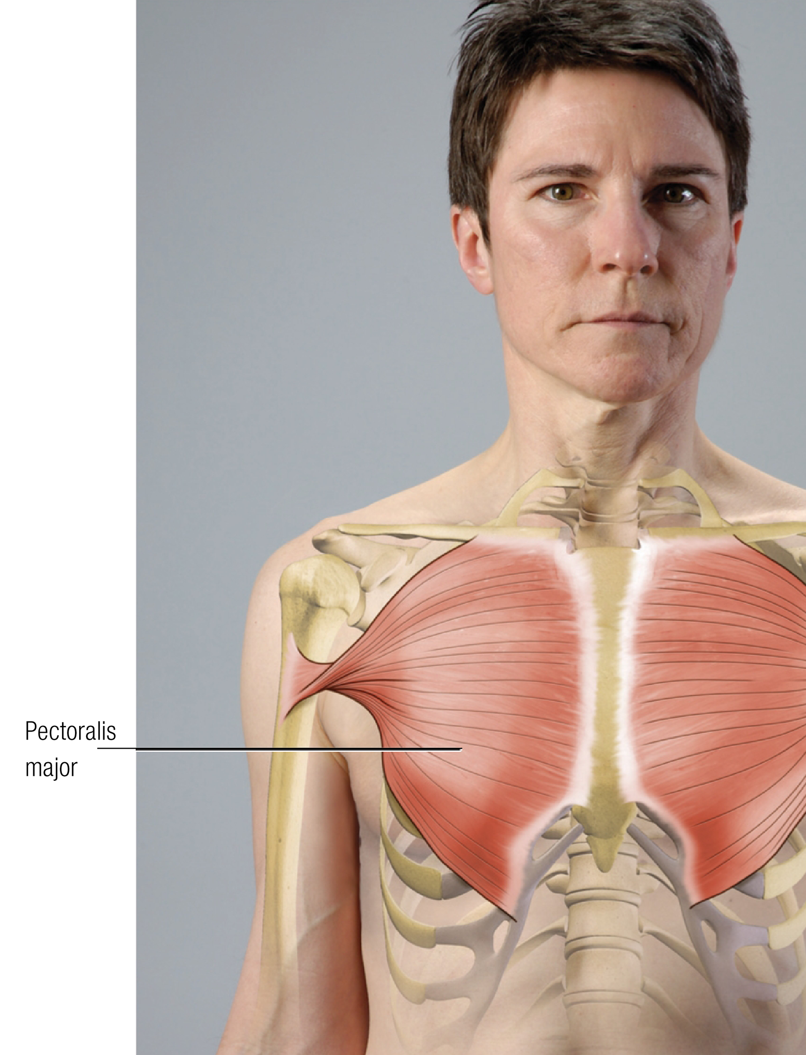
Attachments
Actions
Innervation
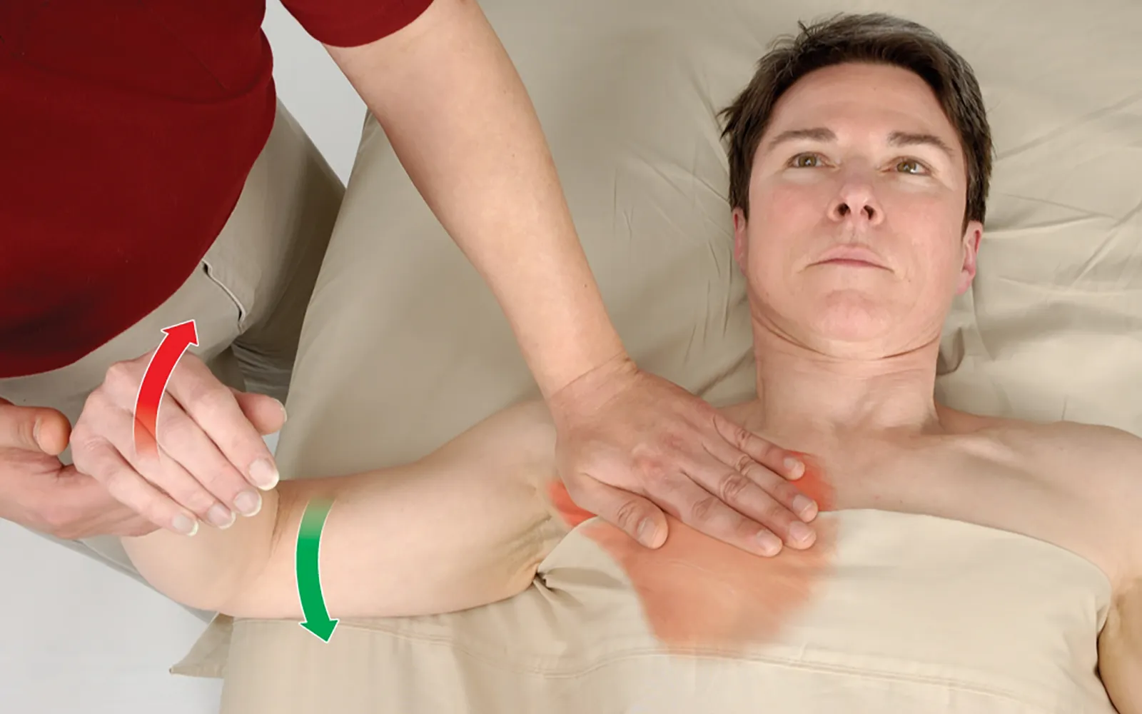
Positioning: client supine with the arm at the side.

Editor’s note: The Client Homework element in Functional Anatomy is intended as a take-home resource for clients experiencing issues with the profiled muscle. The stretches identified in Functional Anatomy should not be performed within massage sessions or progressed by massage therapists, in order to comply with state laws and maintain scope of practice.
Understanding how the pericardium relates to breathing can in turn help clients relax into their massage session.
Superficial fascia may not get the same attention as deep fascia, but it plays an extremely important role in the organization of tissues and the structure of the body.
Understanding tendons—their shapes, lengths, and organization—improves an MT’s touch vocabulary and facilitates a more skilled touch.
While the neck is a bridge, a pathway, the position of the neck and head can also indicate a multitude of other things happening beneath the surface.