

Fascia: The Pericardium-Diaphragm Connection
Understanding how the pericardium relates to breathing can in turn help clients relax into their massage session.
The cartilage in our knees is like the tread on a tire. If the tires are misaligned, the tread wears down unevenly and prematurely. The same thing occurs in the knees. Over time, and with repeated compressive loading, the underlying patellar cartilage and menisci undergo gradual deterioration. Faulty tracking can hasten this process and prematurely wear down vulnerable structures in the knee.
We use the word tracking in two contexts: to describe the actual movement inside the joint, and to describe the clinical skill of observing pathways of movement. Knee-tracking exercises can be used to assess joint mechanics and minimize mechanical stress on the knee during passive movement techniques. In the last column on knee rotations, we tracked axial rotation during knee flexion and terminal rotation during knee extension. In this column, we will look at pathways of motion that occur in two unique structures of the knee: the patella and the menisci.
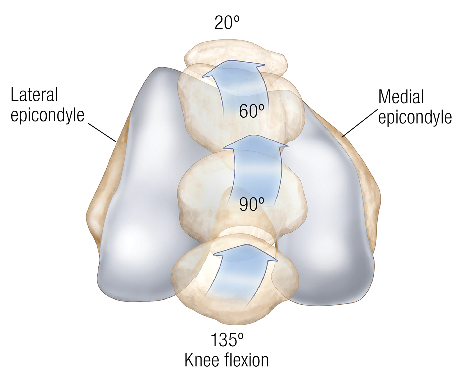
The patella serves as an anatomical pulley to give the quadriceps a corner to pull around. It slides up and down in the intercondylar groove, or notch, along a curved pathway of motion. Thick hyaline cartilage underlying the patella protects the knee from extreme compression and friction generated by contraction of the quadriceps during activities such as walking up or down a hill, which places 200–300 pounds of pressure on the patella. You can track patellar motion by lightly holding the kneecap while slowly flexing and extending the knee.
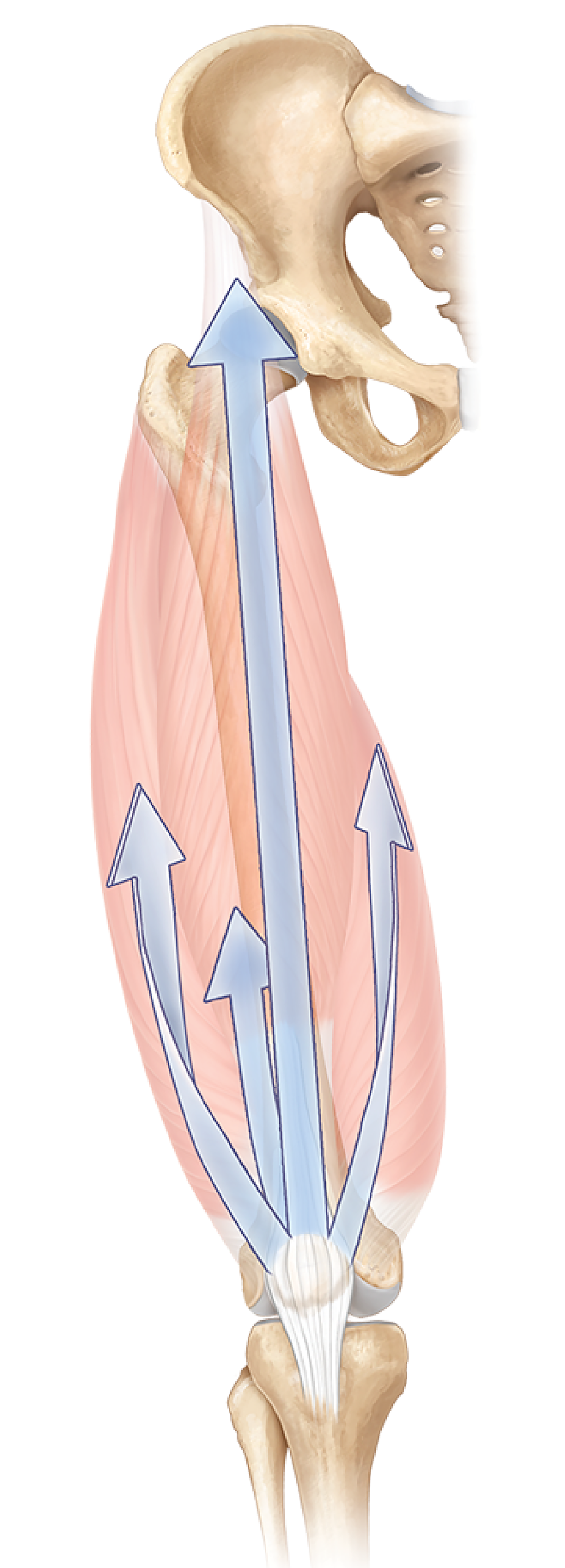
Muscular pulls on the knee must be balanced for optimal patella tracking. Muscular imbalances can trigger inflammatory conditions that produce scarring in patellar cartilage, restricting its gliding motion along the intercondylar groove. For example, when the lateral quadriceps overpowers the medial quadriceps, the patella is pulled laterally off its ideal track. This can lead to the many painful conditions exacerbated by joint motion, such as arthritis and/or the deterioration of patellar cartilage called chondromalacia.
The knee is a double condyloid joint with two distinct sides padded by large, fibrocartilage discs called menisci. The main function of the menisci are to seat and stabilize the femoral condyles to prevent them from sliding or rolling off the horizontal tibial plateau during joint motion. Each meniscus is anchored to the tibia by two ligaments, one at each of its open ends or “horns,” which allows a small amount of motion.
The slight mobility of the menisci allows them to stay under the femoral condyles as the condyles rotate along a migrating axis. Each meniscus moves independently. The lateral meniscus can glide forward and backward twice as far as the medial meniscus, about 12 millimeters. The discs also pivot like cogwheels, counter-rotating in an outward and backward direction during knee flexion and reversing the pathway during extension (Image 3).
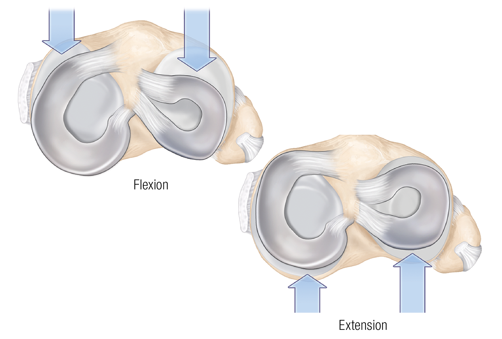
The crushing forces of the condyles deform the menisci, pressing them across the tibia like a pestle pressing a dried apricot across a mortar. If the menisci follow an ideal pathway of motion, they will remain under the condyles throughout range of motion. If a meniscus fails to shift position quickly enough, it can be ruptured or crushed under the pressure of the femoral condyle. A torn meniscus can no longer follow a normal movement range and loses its ability to absorb compressive load.
When working with clients with knee injuries or symptoms, always start by determining if passive knee motion is contraindicated. You can assess patellar and meniscus tracking patterns by palpating these structures as your client moves the knee. Menisci motion is subtle and difficult to detect, but with practice, it can be perceived during palpation. Keep in mind that damaged cartilage is susceptible to reinjury, so when passively moving your client’s knees, make sure to move each joint slowly, with attention to tracking patterns.
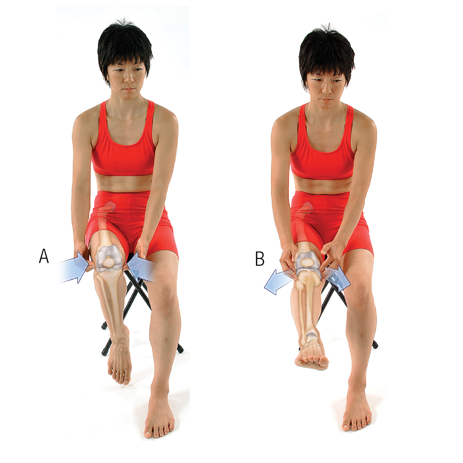
To explore meniscal movement in your own knee (Image 4):
Understanding how the pericardium relates to breathing can in turn help clients relax into their massage session.
Superficial fascia may not get the same attention as deep fascia, but it plays an extremely important role in the organization of tissues and the structure of the body.
Understanding tendons—their shapes, lengths, and organization—improves an MT’s touch vocabulary and facilitates a more skilled touch.
While the neck is a bridge, a pathway, the position of the neck and head can also indicate a multitude of other things happening beneath the surface.