Denise’s Shin Splints

By Whitney Lowe
[Clinical Apps]

Denise has a job in which she sits at her desk most of the day. Fortunately, she has been able to start exercising recently because her company organized a soccer team. Several weeks ago, after their second day of practice, Denise began to experience aching pain in the lower part of her leg. The pain has increased gradually and occurs with each game, continuing for another day or two after the game and then subsiding. The pain is only in her right leg.    
Denise has tried managing the pain with periodic icing and over-the-counter anti-inflammatory medications, but neither approach has been very effective. Her primary interest is to address the pain so she can continue playing soccer.

Key Considerations
First and foremost, it is clear this is a chronic overuse condition that seems directly related to Denise’s recent increase in activity levels. Any time there is a sudden and recent change in activity levels, especially without adequate conditioning, it is a red flag for the development of an overuse disorder. Overuse disorders in the lower leg commonly affect the dorsiflexor muscles, as well as the deep posterior compartment group, so let’s look at the location and function of these two muscle groups in greater detail.

Pain and dysfunction in the dorsiflexor muscle group are generally felt in the anterior shin region and are commonly called shin splints, although that term is used ambiguously. The dorsiflexors play a key role during locomotion and a closer look at their function clarifies why they are so frequently involved in these disorders.

We think of these muscles predominantly as dorsiflexors of the foot, because that is their primary role during their concentric contraction. They also have a key role in decelerating the foot and absorbing impact forces during walking or running. Immediately after a heel strike, the dorsiflexors act eccentrically as the foot moves in plantar flexion to slow the momentum of the foot to keep it from slapping down onto the ground. This repetitive eccentric load at every foot strike is part of what produces the chronic overuse pain of anterior shin splints.

If you are running or walking downhill, the force and effort required to decelerate the foot is even greater. Consequently, you frequently hear of people developing anterior shin splints from a long bout of downhill exercise, like hiking down a mountain. Running also exaggerates the force load these muscles must decelerate. It would certainly fit Denise’s clinical picture for her to have an overuse condition from overloading the dorsiflexor muscles.
Muscles of the deep posterior compartment are also commonly involved in lower leg overuse disorders. The term shin splints can also apply to problems with the posterior compartment muscles, and may be called posterior shin splints or medial tibial stress syndrome (MTSS). Muscles in this compartment include the tibialis posterior, flexor hallucis longus, and flexor digitorum longus (Image 2). The tibialis posterior is most commonly affected, so our investigation will focus on that muscle. However, recent studies also indicate that the soleus muscle attachments can contribute to pain in this region as well.

One of the primary functions of the tibialis posterior muscle is to prevent overpronation in the foot, in which the foot rolls onto the medial side during the weight-bearing phase of a foot strike. It is the eccentric action of the tibialis posterior that prevents overpronation. An excessive wear pattern on the medial side of the shoe’s sole is often evidence of overpronation, as seen in Denise’s case.

The tibialis posterior muscle is difficult to palpate because it is deep in the posterior compartment, which makes investigation of this muscle more challenging. Also, the foot must be everted to stretch the muscle in assessment, but bony structures limit the degree to which the foot can be everted. The tibialis posterior is one muscle that can never be fully stretched as much as it should, so any amount is particularly helpful.  

Now that we have a good understanding of some of the key areas that are likely to be involved, let’s further evaluate the nature of Denise’s primary complaint.

Assessment and Evaluation
Denise originally described the pain as being in the lower leg region, and more medial. This location would tend to implicate problems in the tibialis posterior more than the tibialis anterior. Overuse of the dorsiflexor group tends to cause pain in the upper anterior shin region where the dorsiflexor muscle bellies are located. When pain is felt in the distal lower leg, and especially if it is medial, it is more likely that the deep posterior compartment muscles—or distal portion of the soleus—are involved.

Palpation plays a key role in confirming our suspicions about involvement of the deep posterior compartment muscles. Palpating the medial and distal region of the tibia reproduced some of the characteristic pain that Denise was experiencing, so it was likely we were on the right track. Having our initial assumptions confirmed about the location of pain, we explored further with physical examination.

We had Denise perform a series of foot and ankle movements including dorsiflexion, plantar flexion, inversion, and eversion. She performed the movements first actively, then passively. These evaluations were performed by having her sit on the edge of the treatment table and move her foot in each of the prescribed directions. Following the active and passive movements, we also had her perform resisted dorsiflexion, plantar flexion, inversion, and eversion.
Denise reported no pain with any of the evaluation procedures. This seemed unusual because she was having symptomatic complaints with activity, and it seemed she should have some symptoms when performing some of these motions. When a client has a pain complaint during certain motions but that pain is not reproduced with any of the movement evaluation procedures, then the problem may not be what was initially suspected. However, we couldn’t rule the tibialis posterior out yet because there was another important possibility.

In many cases of musculotendinous injury, the tissue can still be damaged but not cause pain during certain evaluation procedures because not enough stress is placed on the tissue. This is a common pattern with medial tibial stress syndrome, where our standard evaluation procedures do not reproduce the pain sensation because they simply do not stress the tissue enough.

Our solution was to apply moderately deep palpation during these same evaluation procedures. Palpating the tissue, while stressing it through active, passive, or resisted movements, puts significantly more stress on the potentially damaged tissues and can give us more clues about the nature of the problem.

In Denise’s case, pain was slightly reproduced during active movement with palpation, and especially during manual resistance with palpation. The motions that were most painful were active and resisted plantar flexion and inversion. This pattern fit with our suspicion of involvement of the tibialis posterior and/or other deep compartment muscles.
Another way to effectively evaluate many lower extremity disorders is to have the person attempt to do some of the range-of-motion evaluations while in a weight-bearing position. They can’t all be done effectively in a weight-bearing position, but some can. You’ll need to carefully apply your kinesiology knowledge to evaluate which muscles are engaged in concentric or eccentric actions during the various weight-bearing range-of-motion evaluations. For example, if you have the client rise up on tiptoe, that is an active movement that works the plantar flexor muscles concentrically. Coming back to a neutral position, the foot is moving into dorsiflexion, but it is the plantar flexor muscles working eccentrically to get back into a neutral position with the foot flat on the ground.

Based on Denise’s history, and our findings in physical examination, it appeared most likely that some type of chronic overuse disorder was aggravating muscles of the deep posterior compartment, and their irritation was the source of her pain. Having formulated a good hypothesis about the nature of the tissue injury, we now turned our attention to whether or not massage was appropriate, and if so, what would be the most effective treatment strategies.

Treatment Considerations
Because this condition appears to revolve around musculotendinous dysfunction, massage treatment approaches are likely to be very helpful in addressing the complaint. We must keep in mind, however, that despite having identified the deep posterior compartment muscles as the primary culprit, very few soft-tissue dysfunctions are the simple result of problems in one structure alone. Consequently, our treatment approach addressed a number of other tissues as well.

Treatment started with the superficial posterior compartment muscles: the gastrocnemius and soleus. Tightness or dysfunction in either of these muscles can contribute to dysfunction in the deeper posterior compartment muscles. The soleus muscle can also be implicated in many conditions of medial tibial stress syndrome. It is helpful to get the gastrocnemius and soleus as pliable as possible in order to access the deep posterior compartment muscles beneath them. Sweeping cross-fiber and compression broadening techniques are particularly helpful for this purpose.  

After our initial work on the gastrocnemius and soleus, we now focused on the deeper muscles. With muscles like the tibialis posterior that are difficult to access, you have to take advantage of alternative methods to effectively treat that area. An excellent way to access the tibialis posterior and other deep compartment muscles is through active engagement techniques. The active engagement techniques for the posterior compartment muscles take advantage of applying indirect pressure to the muscle while it is engaged in an active contraction. Here are a couple of different ways the deep posterior compartment muscles can be addressed.

Traditionally, active engagement techniques are performed by applying some type of broadening technique to the muscle during the concentric contraction. However, the posterior compartment muscles are too small and too far around the back of the tibia to apply a broadening technique during the concentric contraction. A good alternative is to use a static compression technique during the active concentric contraction. This technique works well to help reduce chronic tightness or any myofascial trigger points that may exist in the muscle.

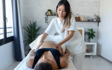
To perform the technique, position the client in either a supine or side-lying position, as long as the foot can move fully through a range of dorsiflexion and plantar flexion. Have the client dorsiflex the foot, place static pressure on the tibialis posterior, and instruct her to plantar flex the foot (Image 4). Pressure is applied during the concentric phase of contraction (plantar flexion). Allow the client to rest and relieve the pressure while she moves the foot back into dorsiflexion. Repeat the process several times until you have worked the full length of the tibialis posterior muscle with static compression during the concentric plantar flexion.

Follow the concentric application with a stripping technique performed during the eccentric or lengthening phase of the muscle. For this technique, have the client in the same side-lying position. You can even have the foot off the end of the table so it can fully move through a range of dorsiflexion and plantar flexion if that works better. Instruct the client to slowly move the foot back and forth (plantar flexion and dorsiflexion). During each dorsiflexion movement, as the tibialis posterior is being elongated, perform a deep stripping technique along the tibial border to the deep posterior compartment muscles (Image 5). Keep in mind that this technique can be painful for the client, especially if a dysfunctional condition like medial tibial stress syndrome exists. However, there are few techniques that are as effective as this in helping to reduce chronic tightness in these muscles and return them to optimal function.

Denise was advised to continue with home-care activities to support the clinical treatment work. Stretching plays an important role in keeping these muscles from experiencing chronic overuse. Because it is difficult to adequately stretch the tibialis posterior muscle, stretching is even more important. In addition to stretching, clients can perform some of the simple active engagement techniques on themselves. The more frequently these muscles are worked, the less they will suffer from cumulative trauma. While the client’s self-massage will not be as effective as what you perform in the treatment room, it is still highly valuable in helping to reduce cumulative stress in these tissues.
It is also a good idea to have the client examine her footwear. Shoes with inadequate support are a frequent contributing problem to chronic overuse problems like medial tibial stress syndrome. Sometimes orthotics in the shoes can be helpful, but this isn’t mandatory and it can be challenging to find an orthotic that is ideally designed for the individual’s unique biomechanical foot dysfunction.

Medial tibial stress syndrome can be effectively treated with massage if the proper applications are used. However, treatment applications are often not specific enough in accessing the deep posterior compartment muscles, and the massage can wind up being ineffective. Following the above protocols should produce effective resolution to the client’s pain.

Whitney Lowe is the author of Orthopedic Assessment in Massage Therapy (Daviau-Scott, 2006) and Orthopedic Massage: Theory and Technique (Mosby, 2009). He teaches advanced clinical massage in seminars, online courses, books, and DVDs. You can find more ideas in Lowe’s free enewsletter—and his books, course offerings, and DVDs—at www.omeri.com.

To read this article in our digital issue, click here.