The Many Faces of Bell's Palsy

By Ruth Werner
[Pathology Perspectives]

Imagine one morning that you wake up and, as usual, shuffle into the bathroom. A quick glance in the mirror yields an unpleasant surprise: somehow in the night the right side of your face seems to have fallen. No matter how hard you try, you can’t seem to make it move. Your right eyebrow sags, and you can’t close that eye all the way. When you try, your right eye rolls up and inward. Your cheek droops, and an attempt at a smile reveals an unfamiliar grimace. Your face doesn’t particularly hurt, but you’re aware of a numb feeling. Furthermore, your taste sensation seems off: everything on the right side of your mouth has a vaguely soapy flavor. Noises are louder too—the running water in the bathroom is enough to make you wince, but only the left side of your face can move. You wonder, as panic begins to rise: Did you have a stroke? Do you have a tumor? Is there any way to fix this? Will you look like this forever?

This is the textbook presentation of Bell’s palsy, which is classified as damage to the facial nerve (cranial nerve VII) leading to a unilateral peripheral facial paresis or paralysis.1 What a diagnosis of Bell’s palsy doesn’t identify is the source of the nerve damage. Viral infection, bacterial infection, mechanical compression, and other factors can all impact the functioning of the facial nerve, but each cause requires a different treatment strategy for the best results. In this article, we will examine the many faces (pun intended) of Bell’s palsy and discuss where and when massage might be included in a treatment strategy.

What is Bell’s Palsy?

Bell’s palsy, named for British physician Dr. Charles Bell, was first described in 1821. It is a sudden onset (usually over the course of 1–3 days, often overnight) of one-sided flaccid paralysis of the face. Flaccid paralysis means the muscles become hypotonic: soft and weak. This is a typical sign of peripheral motor nerve damage.

Because Bell’s palsy comes on so dramatically—often without warning—people fear that they have had a stroke or brain tumor. In the United States, about one out of every 65 people will experience this condition at some point. Pregnant women and people with diabetes are more at risk than the rest of the population, and it is most common among people in their 40s.2

While many experts describe this condition as idiopathic, a source of facial nerve compression can often be found. It is important to identify exactly what is pressing on or irritating the facial nerve, because different problems require different solutions. And while this is usually a self-limiting condition with full or nearly full recovery, appropriate intervention in early stages can speed healing, improve prognosis, and forestall short- and long-term complications.

Anatomy Review

The facial nerve runs a peculiarly convoluted path from its origins on the brainstem to its final destination in the facial muscles and the tongue. It travels through narrow passageways in the cranium, often sharing space with other cranial nerves. It takes at least three sharp turns to get to its destinations. The narrowest tunnel is the temporal facial canal, which allows only about .66 millimeters of clearance. Understandably, any swelling or irritation to the nerve can compress it here, leading to the risk of demyelination and loss of function.

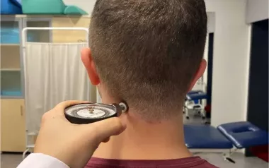
The branches of the facial nerve emerge behind the ear, anterior to the mastoid process. Any bone spur, inflammation, or irritation to the soft tissues at this emergence site can also put mechanical pressure on the delicate nerve tissue. 

Signs and Symptoms

Bell’s palsy has a classic set of symptoms that set it apart from other cranial nerve disturbances; a sudden onset of unilateral weakness of facial muscles is the key sign. This weakness involves the upper as well as the lower face: a feature that is important to distinguish Bell’s palsy from other disorders. Some fibers of the facial nerve also supply the taste buds, salivary glands, and tear ducts, so distorted taste, as well as problems with tears and salivating may also occur. This is especially inconvenient because the muscles of the mouth are weakened, so eating and drinking neatly can become complicated.

The muscles that control eyelids are weak with this condition, so it is difficult or impossible to fully close the eye. Bell’s phenomenon, where the eye rolls superiorly and medially during the attempt to close the lid, is frequent. Sensitivity to light, dryness, dust, and other irritants can be problematic without a functioning eyelid.

Because this condition involves damage to a nerve that has mostly motor rather than sensory neurons, Bell’s palsy tends not to be acutely painful. Many patients report hyperacusis, however. This is the amplification—sometimes painfully so—of sound signals related to the paralysis of the stapedius muscle inside the ear. Chronic headaches and an aching pain at the mastoid process are also common. These pain sensations are quite different from the gripping, seizing, hot-poker-in-the-eye kind of sensations common to trigeminal neuralgia—a different disorder that causes damage to the trigeminal nerve, a mainly sensory nerve that shares some pathways with the facial nerve.

Possible Causes and Treatment Options

Bell’s palsy is a general term that simply describes irritation and or damage to the facial nerve. Finding the source of that damage is important in discerning the best possible treatment options.

Facial nerve irritation is usually brought about by one of two factors: viral infection or bacterial infection. Obviously, these require two quite different treatment strategies, because in addition to taking pressure off the nerve as quickly as possible, it is important to combat whatever pathogens might be attacking the delicate tissue.

Viral Infections
Herpes Simplex

Herpes simplex type 1 is probably the most frequent trigger for Bell’s palsy. This viral infection causes the characteristic lesions we sometimes call fever blisters or cold sores, but in this case the concurrent inflammation also irritates the facial nerve.

Varicella Zoster

This viral infection usually has its first manifestation as chicken pox. Then, the virus goes into hiding, sometimes in the geniculate ganglion of the facial nerve. If the virus reactivates, it causes blisters on the affected neurons: the lesions are sometimes called shingles. When the facial nerve is involved, facial paralysis can occur along with the characteristic pain for which shingles is famous. Facial paralysis along with a varicella zoster outbreak is sometimes called Ramsay-Hunt syndrome.

Other Viruses

Other viruses that can affect the facial nerve include HIV; hepatitis A, B, and C; influenza; Epstein-Barr virus; and cytomegalovirus.3 Viral infections are typically treated with a combination of antiviral drugs (acyclovir, famcyclovir, or valocyclovir), and prednisone—a steroidal anti-inflammatory. If treatment begins within three days of onset of a viral attack, the prognosis for recovery and freedom from complications are good.4

Bacterial Infections

The most common bacterial infection associated with facial nerve damage is from the pathogen Borrelia burgdorferi, the spirochete that causes Lyme disease. When facial paralysis is accompanied by a bull’s-eye rash, or any swelling and redness of the face, a test for Lyme disease should be conducted. In fact, in parts of the country where Lyme disease is endemic, any person with facial paralysis may be tested, just in case.5 Obviously, facial paralysis due to a bacterial infection won’t respond to antiviral medication. This is why it is important to get an accurate diagnosis as quickly as possible so that the correct antibiotics can be used in the hopes of a speedy and uncomplicated recovery.

The same mycoplasmas that cause pneumonia may also irritate the facial nerve.

Other Causes

The list of other possible sources of irritation to the facial nerve is long, and an accurate diagnosis must rule these out. These differentials can include genetic predisposition, tumors of the salivary glands or other tissues, Guillain-Barré syndrome, multiple sclerosis, sarcoidosis, HIV infection, HTLV-1 infection, and barotrauma (damage to tissues related to diving). Fortunately, most of these conditions have a much slower onset than a typical Bell’s palsy case, or they are associated with several other tissue dysfunctions as well, where Bell’s palsy is limited strictly to the facial nerve.

Possible Complications

The most important short-term complication of Bell’s palsy is the risk of damage to the eye, because the facial muscles cannot close the eyelid completely. Patients may be counseled to sleep with an eye patch to avoid scratching the eye. While tear production is impaired and patients need to supplement with moistening drops frequently, many people also have the experience of excessive tearing, which shows how the muscles that control the eyelid have been weakened.

Once the first few weeks have passed and the affected eye is safe, Bell’s palsy usually has an excellent prognosis. Recovery generally occurs within three weeks to three months, but it can take longer. Up to 85 percent of all people experience full or nearly full recovery within a year of onset. About 10 percent have some level of permanent asymmetry in nerve function, and 5 percent have permanent, severe damage.6

Of those who don’t fully recover, their long-term consequences can take several shapes. Incomplete motor recovery may mean that the facial muscles never become fully functional again. One person described it this way: “My dad had Bell’s palsy. He improved over time, but his eye still teared up on him, and he never did get his whistle back.”

In other cases, a person may experience sensory distortion involving the taste buds supplied by the facial nerve. A common complaint: “Everything tastes like soapy beer!”7

A rare but important complication of Bell’s palsy is called synkinesis. Literally this translates to with movement, but in this situation it refers to a malfunction in how the motor neurons of the facial nerve regenerate. A person with synkinesis may experience faulty motor functioning, specifically of the tear ducts and the salivary glands. When he smells something delicious, instead of having his mouth water, he may drip tears. When he is hurt or sad, he may salivate instead of weeping.

And finally, the unopposed muscles on the contralateral side of the face may become chronically tight or even spasmodic. The term hyperkinesis describes the tendency toward spasm that long-term Bell’s palsy patients sometimes experience. Injections with Botolinum toxin are sometimes recommended to temporarily paralyze these overactive muscles.8

Perhaps the most profound effect of Bell’s palsy is on a person’s self-image. One massage therapist describes this client: “I find that what bothers people with Bell’s palsy the most is their appearance. One of my clients was a particularly beautiful woman who was so mortified at the way her face looked that she basically became a hermit until it went away. She started getting house calls instead of coming to the office, she sent her husband or teenagers to do every errand, she pulled a hat way down over her face when she had a doctor’s appointment or something she couldn’t get out of. It lasted for a couple of months or so, and she had been a very social person, too. Basically she let Bell’s palsy take away her whole life while she had it.”

What About Massage?

Bell’s palsy treatment protocols often include massage along with electrical stimulation, exercises, and biofeedback. The general goal with these strategies is to maintain the health and function of the facial muscles while the nerve heals. Research indicates that massage may be helpful in this setting, but no large scale or specifically massage-targeted studies have yet been published.9

Because facial sensation is intact with Bell’s palsy, massage within pain tolerance is probably not only safe, but an important intervention to keep flaccid muscles elastic and well nourished. Massage can specifically stretch and, with client participation, exercise facial muscles that control the eyebrows and mouth for the best possible outcome.

 Ruth Werner is a writer and educator who teaches several courses at the Myotherapy College of Utah and is approved by the NCBTMB as a provider of continuing education. She wrote A Massage Therapist’s Guide to Pathology (Lippincott Williams & Wilkins, 2009), now in its fourth edition, which is used in massage schools worldwide. Werner is available at www.ruthwerner.com or wernerworkshops@ruthwerner.com.

Notes

1.    B. Lo, “Bell Palsy,” Medscape. Available at http://emedicine.medscape.com/article/791311-overview (accessed May 2009).

2.   J. Tiemstra and N. Khatkhate,” Bell’s Palsy: Diagnosis and Management,” American Academy of Family Physicians (2007). Available at  www.aafp.org/afp/20071001/997.html (accessed May 2009).

3.   D.G. James, “All That Palsies is Not Bell’s,” Journal of the Royal Society of Medicine 89 (1996): 184–87. Available at www.pubmedcentral.nih.gov/articlerender.fcgi?artid=1295731 (accessed May 2009).

4.   N. Holland and G. Weiner, “Recent Developments in Bell’s Palsy,” British Medical Journal 329 (2004): 553–7. Available at www.bmj.com/cgi/content/full/329/7465/553?etoc (accessed May 2009).

5.   D.P. Markby, “Lyme Disease Facial Palsy: Differentiation From Bell’s Palsy,” British Medical Journal 299 (1989): 605–6. Available at www.pubmedcentral.nih.gov/picrender.fcgi?artid=1837468&blobtype=pdf (accessed May 2009).

6.   B. Lo, “Bell Palsy.”

7.   Ruth Werner, A Massage Therapist’s Guide to Pathology (Baltimore: Lippincott Williams & Wilkins, 2009).

8.   T.S. Shafshak, “The Treatment of Facial Palsy From the Point of View of Physical and Rehabilitative Medicine,” Eura Medicophys 42 (2006): 41–7. Available at www.ncbi.nlm.nih.gov/pubmed/16565685 (accessed May 2009).

9. Ibid.