Rectus Capitis Posterior Minor

By Christy Cael
[Functional Anatomy]

The rectus capitis posterior minor is one of four pairs of muscles that make up the suboccipitial group (sub meaning “below,” occipital referring to “the occipital bone”). The other three suboccipital muscles include the rectus capitis posterior major, obliquus capitis superior, and obliquus capitis inferior.

Located deep between the base of the skull and first two cervical vertebrae, these muscles maintain alignment between the occiput and upper cervical vertebrae. This supports proper function of associated nervous and circulatory structures. The suboccipitals also generate fine movements of the head, like when you read or scan the road as you walk or drive. These subtle movements are especially important in maintaining spatial orientation and awareness of head position while the body is in motion.

Rectus capitis posterior minor lies at an oblique angle and extends between the first cervical vertebra and the occiput. This muscle is mainly postural and helps stabilize the atlantooccipital and atlantoaxial joints. It also helps maintain the relative position of the upper vertebral foramen to the foramen magnum. Proper alignment in this region supports the flow of blood and cerebrospinal fluid—fluid that cushions and nourishes the brain and spinal cord—to and from the skull. Tension, adhesions, or shortening of the suboccipital muscles can lead to global cognitive dysfunction if circulation or flow of the cerebrospinal fluid is disrupted.

Of the four pairs of suboccipital muscles, rectus capitis posterior minor is the most medial. Its fibers extend directly into the dura—the covering around the brain and spinal cord that contains the cerebrospinal fluid—at the foramen magnum. This dural attachment creates a direct mechanical link between health of rectus capitis posterior minor and the flow of cerebrospinal fluid between the cranium and spinal column. Functional imbalances, hypertonicity, or weakness in this muscle or associated muscles can lead to headaches, cognitive difficulties, and pain.

Palpating Rectus Capitis Posterior Minor

Positioning: client supine

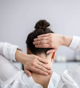
1. Sitting, place both hands palm-up under the client’s head. Find the external occipital protuberance with your fingertips.

2. Slide fingertips caudally and laterally into the suboccipital region near the lamina groove.

3. Curl fingertips upward as the client tucks the chin to slack superficial structures.

4. The client looks up gently to assure proper location.


Christy Cael is a licensed massage therapist, certified athletic trainer, and certified strength and conditioning specialist. Her private practice focuses on injury treatment, biomechanical analysis, craniosacral therapy, and massage for clients with neurological issues. She is the author of Functional Anatomy: Kinesiology and Palpation for Manual Therapists (Lippincott Williams & Wilkins, 2009). Contact her at functionalbook@hotmail.com.