Cool Muscles

Confessions of an Anatomy Geek

By Dr. Joe Muscolino
[Feature]

Key Point
• Individual muscles should be explored for what makes them distinct and extraordinary, in addition to their relationship to the kinematic function of the body.

The myofascial system is an incredible interweaving of tissues, the entirety of which is much greater than the sum of its parts. An analogy might be that the big picture of a jigsaw puzzle cannot be seen and appreciated until all the pieces of the puzzle are placed together in their proper relationship. In this light, each individual muscle functions as part of myofascial meridians and kinematic chains throughout the body. But each muscle also has its own role within this larger scope; therefore, there is value to explore and examine individual muscles. 
But which muscles to choose? Although each muscle is important to the kinematic function of the body, there are some muscles that stand out—muscles that, as an anatomy geek, I can only describe as cool. What makes them cool? They each have something that is distinctive; something extraordinary only that particular muscle possesses. 
Here, I offer an exploration of nine muscles on my cool list (and you’ll find 11 more in thedigital version of this magazine at massageandbodyworkdigital.com). I am sure many of the ones I have chosen would also be on your list. After all, who would not put the piriformis or psoas major on this list, right? But I will also cover a few muscles that might be underappreciated—those that on deeper examination also rightfully deserve the designation of being cool.

1. Piriformis

Let’s start with one of the perennial favorites, the piriformis. What makes the piriformis cool? To begin, the piriformis is the only member of the deep lateral rotator muscle group (along with the superior and inferior gemellus, the obturator internus and externus, and the quadratus femoris) that also crosses the sacroiliac joint (SIJ), and therefore plays a role in SIJ function and dysfunction. In fact, it is often stated that the piriformis is the only muscle that directly crosses the SIJ. This is technically not true because the coccygeus and the superior deeper fibers of the gluteus maximus also cross the SIJ. But the piriformis is likely the most important of these muscles, and it is incredibly important for stabilization of the SIJ. As a result, the piriformis is often overused and tight when there is SIJ dysfunction; and similarly, when the piriformis becomes tight, it can then lead to hypomobility dysfunction of the SIJ. 
The piriformis has another feature that makes it stand out. The sciatic nerve emerges from the internal pelvis through the greater sciatic foramen between the piriformis and superior gemellus into the gluteal region. However, there is a common anomaly in which all, or part, of the sciatic nerve (usually the common fibular nerve portion) emerges either through the belly of the piriformis or superior to the piriformis. The common narrative is that this anomaly can lead to compression of the sciatic nerve, causing sciatica, mimicking sciatic nerve compression by a pathologic disc. However, it would seem likely that a tight piriformis could cause sciatic nerve compression, regardless of whether there is a normal presentation of the nerve or the anomalous presentation. 

2. Sternocleidomastoid

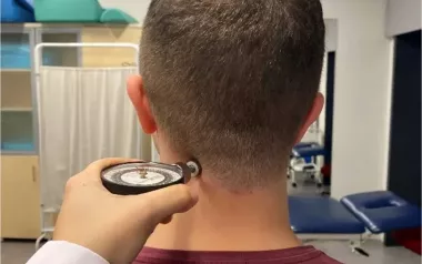
I include the sternocleidomastoid (SCM) in our group of cool muscles because of the sides of the body where it is located. Most muscles of the body cross joints on one side or the other. For example, when looking at sagittal-plane function, flexors of the neck cross the cervical spine in front; extensors cross the cervical spine in back. But the SCM crosses in front and in back. It begins anteriorly on the sternum and clavicle (as its name implies); therefore, as it ascends the neck, it crosses the cervical spine anteriorly. Its path, however, is not perfectly superior. Rather, it is superior and posterior, ultimately attaching to the mastoid process of the temporal bone (also implied in its name); therefore, the SCM crosses the upper cervical spine posteriorly. Due to its path, the SCM flexes the lower and middle neck but extends the head and upper neck (exactly where this division occurs depends on the posture of the person’s cervical spine). Consequently, this gives the SCM a different role than the other neck flexors, such as the scalene and longus muscles. And, as a result, forward-head posture (which involves an overly flexed lower/middle neck with hyperextension of the head) and tight (locked short/overly facilitated) SCM muscles are intimately involved with each other. 

3. Sartorius

The sartorius is well known by many therapists as being the longest muscle in the human body. It is also known for having a name that implies its joint actions. The name sartorius comes from the Latin word for tailor. Before the advent of sewing machines, tailors would sit in a cross-legged position. To attain this position, the thigh (from anatomic position) would have to be flexed, abducted, and laterally rotated at the hip joint, and the (lower) leg would have to be flexed at the knee joint . . . precisely the four major joint actions of the sartorius.  
However, there is another aspect to the sartorius that makes it cool, similar in reasoning to the inclusion of SCM on this list, but even more dramatic. The sartorius attaches proximally on the anterior side of the pelvic bone, travels distally to cross the knee joint posteriorly, but then returns to the anterior side of the body for its distal attachment onto the tibia. No other muscle in the human body has such an unusual path. The natural question is why? The answer lies in evolution. If we look at many quadrupeds (e.g., dogs, cats) who have a flexed position to their back legs, we see that the sartorius travels in a straight line. But when humans stood up to be bipedal, the fascia that held the sartorius in place continued to hold the fascia down against the body such that it now travels from anterior to posterior, and then back to anterior again. Cool! 

4. Brachialis

The brachialis is a simple muscle. It attaches proximally onto the humerus and distally onto the ulna. In doing so, it crosses the elbow joint anteriorly. And because the elbow joint is a uniaxial hinge joint, the action of the brachialis is simply flexion at the elbow joint, nothing more. For this reason, I love to start teaching muscle function by using the brachialis. Simple and elegant. And this is part of its coolness. But much of what makes me love the brachialis so much is its location, which (from an anterior perspective) is deep to the much-more-famous biceps brachii. Because the biceps brachii is more superficial, it gets most of the fame. But the brachialis is usually considered to be the stronger prime mover of elbow joint flexion. I always like to say: “Behind every great biceps brachii is a great brachialis.” (Note—the more superficial gastrocnemius and the deeper soleus of the lower extremity have a similar relationship.)  

5. Diaphragm

The diaphragm is cool for so many reasons. First, it is the primary muscle of respiration. Although there are many accessory muscles of respiration that create forceful inspiration or expiration, contraction of the diaphragm is the only muscle needed for quiet inspiration, and relaxation of the diaphragm is sufficient for quiet expiration. Further, the diaphragm is under both conscious and subconscious control by the nervous system. We can “will” it to contract upon demand, but if we are sleeping or simply not thinking about it, subconscious nervous system control of the diaphragm will maintain its rhythmic contraction. 
It is also cool how diaphragm function can create both abdominal (belly) and thoracic (chest) breathing. When its upper dome moves down toward the rib cage attachment, we have belly breathing; when its rib cage attachment moves up toward the upper dome, we have chest breathing. Finally, the diaphragm is distinctive in that it is usually described as being one muscle, but it truly is two muscles that join at the midline so there actually are right-side and left-side diaphragm muscles, as evidenced by by two separate (right-side and left-side) phrenic nerves. 

6. Transversus Abdominis 

The transversus abdominis is a favorite muscle of Pilates instructors, and indeed all professionals who work with core stability, because it is one of the most important muscles of the core of the body—the powerhouse. The transversus abdominis, as its name implies, runs transversely across the abdomen of the body, in the anterior, lateral, and posterior abdominal walls. So, bilaterally, the two transversus abdominis muscles wrap around the entirety of the abdominal/lumbar region. Even though the transversus abdominis is a skeletal muscle, it actually has no skeletal joint action per se; in other words, it does not actually move a bone at a joint. Rather, its function is to pull in on the abdomen, tautening and stabilizing the abdominal/lumbar region. 

7. Plantaris

The plantaris is a muscle with a lot of personality. I like to describe it as “the little muscle that can.” It attaches proximally onto the femur, and then has a short belly that is only 2–3 inches long that crosses the knee joint posteriorly; it has a tremendously long ribbon-like tendon that travels the entire distance of the (lower) leg to cross the ankle joint and attach onto the calcaneus, right next to the calcaneal (Achilles) tendon. It is a small muscle from the distal femur that has the determination to make its way all the way down to the foot! A lot of pluck! 

8. Extensor Carpi Ulnaris 

I include the extensor carpi ulnaris (ECU) on this list for an unusual reason—because manual/massage therapists so often skip this muscle when working on clients. Therapists usually work the extensor compartment of the forearm with the client lying supine and the forearm pronated. But the ECU is situated right next to the ulna, a bit around the bend so to speak, so therapists usually do an excellent job of working the rest of the musculature of the extensor compartment, but often miss the ECU itself. This is a pity because the ECU is often tight. It is a cool muscle deserving of manual therapy treatment.

9. Psoas Major

For our final muscle under consideration, we have the psoas major, likely the most controversial muscle in the human body. Limiting our discussion to the sagittal plane, the psoas major is an important hip flexor that, when tight, can exert its (reverse-action/closed-chain) pull upon the pelvis, thereby increasing the anterior tilt of the pelvis. This changes what is known as the sacral-base angle, resulting in an increased lordotic curve of the lumbar spine, which then kinematically influences the sagittal-plane posture of the rest of the spine above. Therefore, through its pull on the pelvis, the psoas major, like all hip flexors, can indirectly affect spinal posture. 
But the psoas major also crosses spinal joints directly, attaching as superiorly as T12, and can therefore directly influence spinal posture. Again, looking at the sagittal plane, the line of pull of the psoas major crosses the lumbar spine anteriorly, so it should flex the spine. For this reason, there is an argument made that sit-ups, especially the old-fashioned ones with the hips and knees straight, disproportionately strengthen and tighten the psoas major, which then increases anterior pelvic tilt, thereby increasing lumbar lordosis. For this reason, one paradox of psoas major function is that even though it is a spinal flexor, it can result in increased lumbar extension (lordosis is extension). 
However, there is another level to the paradox of psoas major function that results in increased lumbar lordosis; its line of pull relative to the mediolateral sagittal-plane axis of motion for the lumbar spine can change with changes in posture of the spine. In typical anatomic position posture, the psoas major crosses the lumbar spine anteriorly. But if a person has a hyperlordotic lumbar spine, the line of pull of the psoas major moves posteriorly to cross the lumbar spine (especially the upper lumbar spine) posteriorly, thereby becoming an extensor of the spine. This adds to the paradox that is the psoas major. It can be both a flexor of the lumbar spine and an extensor. 
A third aspect of the psoas major that makes it so cool is that palpation and massage of the psoas major, which is technically a muscle of the posterior abdominal wall, is done from the anterior perspective by sinking in through the abdominal contents to arrive at the psoas major against the spine. Although the psoas major is most often palpated and worked with the client supine, it can also be worked with the client sidelying, three-quarter sidelying, seated, standing, and prone.

10. Extensor Carpi Radialis Longus and Extensor Carpi Radialis Brevis 

The extensor carpi radialis longus (ECRL) and extensor carpi radialis brevis (ECRB) are cool because even though these extensors cross the wrist joint posteriorly, they actually cross the elbow joint anteriorly as part of the radial group of muscles (along with the brachioradialis), so they flex the elbow joint. This is where close attention to the name of a muscle matters. Extensor carpi means “extension of the wrist,” but it does not necessarily mean they are also extensors of the elbow joint. This distinction matters when we are engaging the muscles during palpation assessment, and also when we look to stretch the muscles. The ECRB is also important because it is one of the principal muscles involved with tennis elbow (lateral elbow tendinopathy). 

11. Flexor Digitorum Superficialis and Flexor Digitorum Profundus

Viewed together, the flexor digitorum superficialis (FDS) and flexor digitorum profundus (FDP) muscles of the upper extremity have an extremely simple yet profound presentation to their structure. The FDS attaches distally onto the middle phalanges of fingers 2–5 (index through little) of the hand; the FDP attaches onto the distal phalanges of these fingers. However, as the names imply, the FDP is deeper than the FDS. So how can the FDP make its way to the distal phalanx of a finger if the FDS stops at the middle phalanx and blocks its path? The answer is so simple, yet so elegant. The more superficial FDS tendon splits to attach to the sides of the middle phalanx so that the deeper FDP can make its way through the split FDS tendon to continue on to the distal phalanx of that finger. Cool! 

12. Flexor Digitorum Brevis and Flexor Digitorum Longus 

In the lower extremity, the relationship between the more superficial flexor digitorum brevis (FDB) and the deeper flexor digitorum longus (FDL) mirrors that of the flexors digitorum superficialis and profundus of the upper extremity. The superficial FDB attaches onto the middle phalanges of toes 2–5 but does so by splitting to allow the deeper FDL passage to the distal phalanges of those toes. 

13. Quadratus Plantae

The quadratus plantae involves what I consider to be the simplest, yet coolest aspect of muscle function in the entire human body—its relationship with the flexor digitorum longus (FDL) muscle. The FDL attaches proximally in the leg and then travels distally to attach onto the distal phalanges of toes 2–5. As its name implies, the FDL flexes toes 2–5.

The problem is that any extrinsic muscle of the foot that enters the foot from the leg cannot cross dead center over the calcaneus because it would be compressed or crushed with weight-bearing. So, extrinsic foot muscles must enter the foot on either the medial or the lateral side. The FDL enters on the medial side, posterior to the medial malleolus of the tibia. The problem with this is that its line of pull on the toes will not be perfectly anterior to posterior; therefore, it would not perfectly flex the toes. Instead, it would splay the toes toward the medial side of the foot. This, in time, would cause damage to the uniaxial interphalangeal joints of the toes. What is the solution? We need an accessory muscle to the FDL that can straighten out its line of pull. The quadratus plantae accomplishes this. It attaches into the distal tendon of the FDL, and when the FDL contracts, the quadratus plantae contracts, contributing a more lateral component to the FDL’s line of pull. Ingenious! Ironically, the old name for quadratus plantae was flexor digitorum accessorius, which I believe was a better name. It told us of its simple yet profound contribution to the FDL. Quadratus plantae: Cool! 

14. Adductor Magnus

I believe the adductor magnus is the most underappreciated muscle in the human body. As its name implies, it is a tremendously large muscle. Yet, the immenseness of its size cannot be appreciated from superficial anterior or superficial posterior views because it is deep from both perspectives. It can be viewed as a shelf that the more anterior muscles of the adductor group sit on, and it can be viewed as a shelf that the medial hamstrings sit on. To understand this muscle you must either look at a medial view of the thigh, wherein at least a small amount of it is superficial and shows, or even better, look at deeper views from the anterior or posterior perspective. 

But being large is not enough to qualify it to be on our list of cool muscles. So why do I include adductor magnus here? One reason is that it has a twist in its fibers, similar to the latissimus dorsi and the levator scapulae. But even more fascinating is that it is a transitional muscle between the medial and posterior compartments of the thigh. The adductor magnus has two heads. The anterior head is innervated by the obturator nerve, as are the majority of the adductor muscles. But the posterior head is innervated by the sciatic nerve, as are the hamstrings. And the posterior head attaches proximally onto the ischial tuberosity (like the hamstrings), and extends the thigh at the hip joint (like the hamstrings). For these reasons, the adductor magnus is often referred to as the fourth hamstring, although this designation should really be applied only to its posterior head. An immense adductor muscle that has a twist and is an honorary member of the hamstring group . . . that is cool!

15. Gluteus Medius

While we are in the region of the hip joint, let’s discuss the gluteus medius for a moment. I like to call the gluteus medius the deltoid of the hip. This is because, like the deltoid, the gluteus medius has anterior, middle, and posterior fibers. And these fibers act upon the thigh at the hip joint in the same manner that the deltoid acts upon the arm at the shoulder joint. Like the anterior deltoid, the anterior gluteus medius flexes, abducts, and medially rotates. Like the middle deltoid, the middle gluteus medius effectively does pure abduction. And like the posterior deltoid, the posterior gluteus medius extends, abducts, and laterally rotates. (By the way, the gluteus minimus could be similarly described.)

But another fascinating aspect of the gluteus medius is that its major function is its reverse-action/closed-chain pull upon the pelvic bone toward depression. A pull toward pelvic depression acts to stabilize the pelvis by preventing the opposite-side pelvic bone from depressing when the opposite-side foot leaves the ground during its swing phase of the gait cycle (walking). Weakness of the gluteus medius causes a dysfunction when walking, known as Trendelenburg gait (a falling of the pelvis toward the swing-leg side, similar to how models walk). It could be noted that the reverse/closed-chain function of the deltoid at the shoulder joint is also important because it acts to stabilize (and/or downwardly rotate) the scapula when the arm raises into abduction and/or flexion (scapulohumeral rhythm). 

The gluteus medius is underappreciated in its size because it is next to the largest muscle in the human body—the gluteus maximus. But the gluteus medius is, in its own right, a tremendously large and cool muscle. 

16. Obturator Internus

Staying with the theme of the hip joint, we have the obturator internus, which is another member of the deep lateral rotator group. What makes the obturator internus so interesting is that it turns around a fulcrum created by the pelvic bone that changes its line of pull by 90 degrees. Very few muscles in the human body change their line of pull so dramatically. Cool!

17. Coccygeus

The coccygeus is a pelvic floor muscle, but one that can be easily accessed and worked with massage. Most pelvic floor musculature is the province of physical therapists and manual therapy physicians who are licensed to work the pelvic floor by entering a body orifice (vagina or anus). However, the coccygeus is easily accessible for manual therapy from the outside. A further aspect that qualifies this muscle as cool is that its line of pull actually crosses the sacroiliac joint (SIJ) and can therefore have a role in SIJ function and dysfunction. The coccygeus lies directly inferior to the piriformis and attaches from the ischial spine to the coccyx and sacrum. In fact, it attaches just as much to the sacrum as it does onto its eponymous coccyx. I believe that if this muscle had sacrum in its name (perhaps ischiosacralis or something similar), it would be viewed as a much more important and massage-friendly muscle by therapists. 

18. TERES MAJOR

The teres major falls into the category of underappreciated muscles because it is linked with the latissimus dorsi. The teres major lies next to the latissimus dorsi in the posterior axillary fold of tissue. Like the latissimus dorsi, it starts posterior and ends anterior, and shares the same shoulder-joint functions of extension, adduction, and medial rotation. For this reason, the teres major is sometimes referred to as the little helper or little brother to the latissimus dorsi. But there is nothing little about the teres major. In fact, in the posterior axillary fold of tissue, the teres major is larger than the latissimus dorsi.

Another reason the size and strength of the teres major are underappreciated is that it attaches to only the lower one-third of the lateral border of the scapula, whereas the teres minor has a broader attachment to the upper two-thirds of the lateral border. Even though the teres major has a smaller expanse of scapular attachment, it is a much larger muscle than the teres minor. The teres major is a large and tremendously strong muscle, and extremely important with shoulder joint function. 

19. Semispinalis Capitis

The semispinalis capitis is another underappreciated muscle. It is the largest muscle in the posterior neck, but because it lies deep to the upper trapezius, it is often overlooked. In fact, it is common for a tight semispinalis capitis (as well as the deeper semispinalis cervicis) to be mistakenly assessed as a tight upper trapezius or perhaps tight erector spinae musculature (even though there is very little erector spinae musculature in the neck). If you ever have the opportunity to attend a cadaver lab, be sure to look at (and, if possible, palpate) the semispinalis capitis muscle. 

20. Fibularis Longus 

The fibularis longus has three interesting aspects to it. First, along with the tibialis anterior, these two muscles comprise the stirrup muscles because they wrap around the foot in such a manner that they form a stirrup that supports the foot. Second, even though the fibularis longus is a muscle of the lateral compartment of the leg, it attaches to the medial side of the foot; it arrives at the medial foot by passing through the deep layer (layer IV) of the plantar side of the foot. Another cool thing about the fibularis longus is that it used to be called the peroneus longus. Its name was changed at an anatomy congress in 1999 because the Latin root for lateral leg, fibularis, is easier for English speakers to learn than the Greek root, peroneus.

So, there you have it—20 muscles that have all earned their place on my cool muscles list. 

 Dr. Joe Muscolino has been a manual and movement therapy educator for more than 35 years. He has created several online streaming subscription platforms for manual therapy continuing education, including LearnMuscles Continuing Education (LMCE) with more than 3,000 video lessons and more than 300 hours of NCBTMB credit. He has also created Muscle Anatomy Master Class (MAMC), Bone and Joint Anatomy Master Class (BAJAMC), Visceral Anatomy Master Class (VMC), and Kinesiology Master Class (KMC). And he is the author of multiple textbooks with Elsevier; and has authored more than 90 articles. For more information on any of Dr. Joe’s content, visit learnmuscles.com. To contact Dr. Joe directly, you can reach him at joseph.e.muscolino@gmail.com.