The Fascial Network

Our Richest Sensory Organ

By Robert Schleip
[Feature]

 Key Points

• The estimated number of nerve endings in the body-wide fascial system is 250 million. Compared with the estimated 200 million nerve endings in the skin, this suggests the human fascial network constitutes, in fact, our richest sensory organ.
• Research shows that emotional stress may exert significant influences on the expression of myofascial pain.

For many decades, and for most Western medicine professionals, fascia was primarily considered an inert wrapping organ, merely giving mechanical support to our muscles and other organs. Yes, there were some early histological reports about the presence of sensory nerves in fascia, but these were largely disregarded and did not affect the prevailing medical understanding of musculoskeletal dynamics.1 While Moshe Feldenkrais and Ida Rolf were apparently not aware of the intriguing importance of fascia as a sensory organ for our body perception, Andrew Taylor Still, the founder of osteopathy, wrote that, “No doubt nerves exist in the fascia . . .” and suggested all fascial tissues should be treated with the same respect as if dealing with “the branch offices of the brain.”2 
In the late 1980s, Jaap van der Wal reported about the rich presence and complex arrangement of sensory nerve endings in the muscular fascia of rats,3 but this was largely ignored in mainstream medicine, at least for the subsequent two decades. What really changed the general medical perspective in a profound manner was the first international Fascia Research Congress, held at the Harvard Medical School Conference Center in Boston in 2007. During the congress, three teams from different universities and countries reported their independent findings of a rich presence of sensory nerves in fascial tissues.4 Following that event, many studies were published about fascial innervation, suggesting that fascia plays an important role in the perception and internal representation of our own bodies. 
More recently, the German neurophysiologist Martin Grunwald estimated there were 100 million nerve endings in the body-wide fascial net.5 This number related only to the mass of dense fibrous connective tissues, which was estimated at 5 kg for an average adult human body. In a subsequent calculation, a new estimate was tabulated using a more functional definition of “the fascial system,” one that was based on recommendations from the Nomenclature Committee of the Fascia Research Society. That definition broadened to include all fibrous collagenous connective tissues, including intramuscular connective tissues, joint and organ capsules, ligaments, aponeuroses, tendons, loose connective tissues, epineural envelopes, as well as the intramuscular septi.6 With this revised definition, the mass of fibrous connective tissue in humans increased to 12.5 kg or 17 percent of the total body weight. Subsequently, the estimated number of nerve endings in the body-wide fascial system increased to 250 million.7 Compared with the estimated 200 million nerve endings in the skin8 (which based on its richness and complexity is commonly described as “a sensory organ”), this new calculation suggests the human fascial network constitutes, in fact, our richest sensory organ.

Different Types of Fascial Sensory Receptors

Based on this recognition of the importance of the fascial system as a sensory organ, practitioners are increasingly interested in understanding which exact sensory qualities are served by this rich innervation. Image 1 demonstrates the most important components. The highest proportion is constituted by interstitial receptors (which includes all neurons terminating in free nerve endings, including C-fibers and A-delta fibers). These neurons are thin and sensitive to mechanical stimulation. If the extracellular matrix is chemically altered, or if the mechanical stimulation is too strong, many of these receptors could also become nociceptors (signaling potential tissue damage to the central nervous system). Interestingly, the mechanical activation threshold of the fascial free nerve endings is two times lower than in skin and muscle.9 In addition, it was demonstrated that the free nerve endings in the human thoracolumbar fascia are significantly more sensitive to chemical irritation compared to the underlying muscles, and that they tend to maintain a long-lasting hypersensitivity.10 
For manual therapy practitioners, it is important to realize that not all interstitial receptors can be classified as nociceptive. For example, some of them are sensory devices for thermoception. Interestingly, in fascial tissues, most of the interstitial (between fibrous tissue) neurons are so-called polymodal receptors, meaning they are responsive to more than one kind of stimulation. 
There are four types of specialized proprioceptive mechanoreceptors in fascia: Golgi, Pacini, Ruffini, and muscle spindle receptors.11 

Golgi Receptors

Golgi endings are slowly adapting receptors that respond to tensional loading. While they were previously assumed to be present only in tendinous tissues, their existence in other fascial tissues has been repeatedly confirmed by several independent investigators.12 Their presence is particularly enriched in the myotendinous junctions and close to the intermuscular septa. Stimulation of Golgi receptors tends to trigger a relaxation response not in the whole body, but only in those skeletal muscle fibers which are directly linked with the respectively tensioned collagen fibers. Nevertheless, if tendinous extramuscular tissues are stretched “in series” (i.e., in contrast to a parallel alignment) with muscle fibers that are in a relaxed condition, then most of the respective elongation will be “swallowed” by the more compliant myofibers. In this way, the respective stretching impulse may not provide sufficient stimulation for eliciting any muscular tonus change.13 
A practical conclusion of this may be that a stretching impulse, aimed at reaching the tendinous tissues, may benefit from including some moments in which the lengthened muscle fibers are actively contracting or are temporarily resisting their overall elongation. In addition, a manual approach in which the fascial tissues are stretch-loaded in a direction orthogonal to the direction of closest myofibers (as is frequently practiced in manual cross-fiber techniques based on the teaching of Tim Bowen), it could well be that sufficient elongation can be achieved in the passively lengthened collagenous fibers, despite the more compliant myofibers in their vicinity. 

Pacini Receptors

The Pacini receptors are rapidly adapting and therefore tend to stop responding to a continuous stimulus. In contrast, they are very sensitive to dynamic changes in mechanical stimulation. Gentle rocking, as well as more rapid vibratory stimulation, appears to be a suitable stimulation method for these receptors. Such treatment could therefore have beneficial effects on proprioception. 

Ruffini Receptors

Ruffini receptors are slowly adapting, and tend to remain sensitive in response to a continuous stimulus. They can monitor longer-lasting postural sensations, as well as slowly melting myofascial manipulation techniques. In addition, they are highly sensitive to differences in directional shear loading, which may correspond to the “local listening” approach used by many osteopaths when they use their hands to detect the specific direction a given tissue “wants to move.” 

Muscle Spindle Receptors

The muscle spindle receptor is a fusiform specialized mechanoreceptor. It includes several intrafusal muscular fibers and is surrounded by a strong capsule of connective tissue.14 While it did not exist in our early evolutionary fish ancestors, it is particularly expressed in muscular regions with a strong antigravity regulation function. Combined with the recent 2021 Nobel Prize in Physiology or Medicine that was surprisingly dedicated to sensory receptors in the human body, increasing attention is currently placed on the newly discovered PIEZO2 membrane receptors on the surface of human muscle spindles. These tiny membrane receptors, several magnitudes smaller than the spindle itself, are highly sensitive to mechanical stimulation and play an important role in proprioception. Associated with genetic variations in our population, it was found that a high proportion of people with adolescent idiopathic scoliosis are characterized by a diminished expression of these PIEZO2 receptors in their muscular tissues and by a related impaired proprioception. For many therapists and researchers dealing with scoliosis, even more exciting was the discovery that these persons express much smaller muscle spindles compared to healthy non-scoliotic persons.15 
Spindle capsules are usually embedded into the intramuscular fascial layer of the perimysium. Since muscle spindles can detect tensional differences of just 3 grams, it is assumed their sensitivity can be significantly impaired by increased stiffness of the surrounding perimysium.16 Such stiffness is frequently associated with chronic immobilization and with many fibrotic pathologies. Such tissue changes may therefore result in impairments of proprioceptive refinement comparable to those associated with genetically associated spindle differences in adolescent idiopathic scoliosis. 

Fascia as a Pain Originator

Most studies about fascial nociception have been directed at the thoracolumbar fascia (or lumbar fascia in some animals). Based on these, it is known that this fascia is densely innervated and its free nerve endings can provide nociceptive signaling to dorsal horn neurons in the spinal cord.17 Interestingly, it was shown that stimulation of this fascia (e.g., via injection of saline solution as a mild irritating substance) triggers stronger and longer-lasting pain sensations compared to stimulation of the muscles underneath.18 Such fascial pain was experienced by the patients as burning, throbbing, and stinging. 
The decreased pain threshold in muscular tissues after intense exercise—described in sports medicine as delayed onset muscle soreness—seems to consist, to a large degree, in a hypersensitivity of the fascial envelopes (epimysia) around the involved muscles. In fact, after experimental induction of delayed onset muscle soreness, pain thresholds of the fascia decreased significantly more than those of the underlying muscle tissue.19 These findings suggest that fascial tissues can play a frequent role in providing nociceptive stimulation (or irritation) to the spinal cord. Whether a nociceptive stimulation leads to pain perception in a specific client depends on many additional factors, including their danger-oriented cortical processing. However, the described findings indicate that the tissue origin often makes a substantial difference, since the central nervous system tends to respond to fascial nociception—in contrast to muscular nociception—with a particularly sensitive pain-associated responsiveness.
Manual practitioners who completed their basic training in neurophysiology in the last century often use the “gait control theory” as an explanatory model for the pain-reducing effect of their work. Recent advances in pain sciences have shown this to be only partially correct.20 Proprioceptive signaling can provide a pain-inhibiting effect, particularly to their specific innervation region at the spinal cord. However, for this effect, the speed of the incoming neural stimulation is apparently not a decisive factor. 
Instead, novel investigations revealed that the synapses of the polymodal receptors in the posterior horn of the spinal cord are eager for any kind of stimulation from their respective peripheral innervation region. They seem to be easily satisfied in case sufficient proprioceptive information is supplied to them from these tissue regions via these polymodal receptors. However, when the connective tissue matrix surrounding the respective sensory receptors is altered, these interstitial neurons tend to actively lower their threshold for nociceptive stimulation, which may then lead to myofascial pain. In addition, they may actively give off cytokines that sensitize polymodal neurons in their peripheral innervation region and predispose these areas to a nociceptive function.21 A seemingly miniscule mechanical stimulation, such as a leg-length difference of only 1 mm, can then lead to a nociceptive response within the intricate network of these intrafascial polymodal receptors. 

Fascial Interoception

The term interoception is usually applied to describe body perceptions that are less concerned with where our body is in space and in relation to gravity, and more with how it is doing in its constant search for homeostasis related to our physiological needs. Interoceptive signaling is therefore associated with somatic perceptions, such as temperature changes, hunger, thirst, nausea, tingling, soreness, oxygen supply, and muscular effort, as well as a sense of belonging (versus alienation) regarding specific body regions.22 The peripheral sensory receptors related to interoception are all free nerve endings—they are interstitial receptors. Most of those receptors are located in visceral connective tissues and constitute an essential part of what is frequently referred to as the enteric brain. In addition, other interoceptive interstitial receptors are located within perimysial and endomysial intramuscular fascial tissues. 
The neural stimulation from those interoceptive nerve endings does not follow the usual afferent pathways toward the somatomotor cortex in the brain; rather, these neurons project to the so-called insular cortex, an area of cortical gray matter that is not located on the brain’s outer surface but is folded down or internally into the depth of the forebrain. In this walnut-sized brain area, perceptions about internal somatic sensations are associated with emotional preferences, expectations, and feelings. Patients with disturbed functioning of the insula may still have full biomechanical functioning and achieve high IQ levels in respective cognitive tests, but they are usually impaired in their social skills and are unable to make reasonable decisions in complex social situations.23
Several health-related impairments, such as low-back pain, scoliosis, or complex regional pain syndrome, are associated with a dysfunctional proprioceptive acuity. In contrast, other health conditions tend to be more clearly associated with dysfunctional interoceptive processing (see Image 2).
These latter conditions include eating disorders, anxiety, depression, irritable bowel syndrome, compulsive disorders, alexithymia (inability to recognize and express one’s own emotional states), and posttraumatic stress disorders. It therefore makes sense that massage and bodywork practitioners carefully examine their habitual preferences in fostering the direction of the suggested somatic curiosity of their clients. A rigid reliance on proprioceptive attention, with questions like “Which of your two shoulders rests more fully on the support of the table underneath you?” may provide limited long-term therapeutic improvements if applied to clients for whom a more interoceptive perceptual refinement approach may be required. In these cases, a skillful fostering of visceral fascial sensations, via gentle visceral manipulation, or a direction of the client’s mindful attention toward potential sensations of streaming or “energy flow,” or temperature changes in a particular body region may sometimes provide more profound effects than the often-habitual focus on musculoskeletal sensations.

Autonomic Nervous System and Fascial Stiffness

Recently, Siegfried Mense from Heidelberg University reported, based on his extensive histological investigations, that the most numerous receptor type in human fascia are sympathetic nerve endings. No other receptor type, whether it is Golgi receptors or substance P positive nociceptive free nerve endings, has such a rich expression in fascial tissues as the sympathetic nervous system, the receptors of which make up approximately 40 percent of the entire fascia innervation.24 
Researchers agree that most of these nerve endings are vasoconstrictors, meaning that they control the flow of blood, including the microcirculation through tiny arterioles and venules. They therefore influence regional changes in tissue temperature, supply of oxygen and other nutrients, and inflow of fresh water into the ground substance and the removal of waste products from a given area. In other words, how warm or cold a tissue region is, and to what degree it may tend to accumulate inflammatory cytokines and free-radicals due to relative stagnation in fluid flow, is largely influenced by the numerous sympathetic nerve endings—approximately 100 million—in our body-wide fascial network. 
The influence of emotions on the autonomic nervous system (ANS), as well as on our immune system, has been the topic of the well-established field of psychoneuroimmunology. Combined with the rich sympathetic control of microcirculation, these insights explain how subtle changes in skin temperature often reflect acute and chronic changes in emotions.25 Psychological researchers therefore tend to use thermography in an increasing manner in their investigations to explore this profound interaction. For manual practitioners, detection of skin temperature changes through a well-trained practitioner’s hand may provide almost equally precise information.26 
Interestingly, not all sympathetic nerve endings in fascia terminate near blood vessels. A significant proportion of them seem to have no association with blood flow regulation, as they are not located near any blood vessel. Neuhuber and Jänig27 assume they have a trophic function, meaning they influence the biochemical milieu around them (see Image 3).
These sympathetic nerve endings might also play a role in the long-term regulation of fascial tonicity. The German physiologist Jochen Staubesand proposed a close connection between chronic sympathetic activation and fascial stiffness when he discovered contractile cells with smooth-muscle cell morphological features in the dense fascia covering the human lower leg, together with a rich presence of sympathetic nerve endings. Based on this finding, he proposed that our nervous system regulates myofascial tonus adjustments via two different tonus adjustment systems. The first system involves rapid contractile changes of skeletal muscle fibers. This is regulated from the somatic nervous system via changes in alpha- and gamma-motor neuronal activation. In parallel to this rapid muscular regulation, the stiffness of the fascial tissue elements is regulated via the ANS, which leads to slower and longer-lasting tonicity changes for which chronic emotional stress and other expressions of psycho-emotional health play significant contributory roles.28 In an interview I conducted with Staubesand in 2003, he said, “It now appears that the fascial tonus might be influenced and regulated by the state of the autonomic nervous system. Plus (and this should have ramifications for your work), any intervention in the fascial system may have an effect on the autonomic nervous system in general and on all the organs which are directly affected by the autonomic nervous system. To put it more simply: Any intervention on the fascia is also an intervention on the autonomic system.”29 
This intriguing new hypothesis motivated our small research group at Ulm University (Germany) to further explore the active contractile capacity of fascial tissues with extensive laboratory investigations for more than 10 years. In our concluding publications, we documented that we found contractile cells in all examined fasciae, although with differences in their density. Interestingly, the human thoracolumbar fascia expressed a richer density of such cells compared to other human fascia or to the lumbar fascia from animals we had explored. The related cells are not smooth muscle cells but are fibroblasts with smooth muscle-like contractile features, called myofibroblasts.30 Such cells have previously been observed in wound healing and in fibrotic pathologies, such as in frozen shoulder or in Dupuytren’s contracture (also called Viking’s hand or palmar fibromatosis). 
When we examined the contractile response of different fascial tissues in response to chemical stimulation (with small tissue pieces suspended in a pre-stretched condition in a physiological solution bath), we found a clear stiffening response to several cytokines of the human body, most notably to thromboxane and to TGF-ß1 (a cytokine known as transforming growth factor beta-1 that is associated with tissue fibrosis and wound healing). The contractile response speed or magnitude was extremely small when viewed in a time frame of minutes or hours. This seemed to fit to the reported slow contraction speed of fibrotic pathologies (like in burn scar injuries or frozen shoulder), which can contract with a speed of approximately 1 cm per month. Staubesand suggested our group explore whether stimulation of fascia with adrenaline or other synaptic transmitter substances of the ANS might be able to induce any contractile response. Against his and our expectations, this was not the case, meaning we were not able to induce a clear stiffening response in our experimental setting in response to adrenaline, acetylcholine, etc. 
Years later, we learned about a new insight in the field of psychoneuroimmunology. In this field, it had been known for a long time that chronic sympathetic stress activation can induce a different activation of T3 immune cells in the lymph nodes. This functional influence is often observed when clients are unusually resistant to infections during the final days of an intense professional deadline but are more likely to catch such infections during the first days of their recovery afterward. For a long time, the missing link or specific cytokine was not known between the sympathetic activation and the immunological response. When the missing link was identified, it was a big surprise, at least for us, as this link is the very cytokine called TGF-ß1, which had triggered the most potent tissue stiffening responses in our experiments.31 
This messenger substance is not a hormone, but a cytokine (a messenger substance produced by cells to communicate with each other) and is created by fibroblasts and then stored in specific pockets of their surrounding ground substance. During times of intense mechanical or biochemical stress, they then open these pockets and use their prefabricated “miracle drink” to mutate into powerful myofibroblasts with a dramatically increased contraction power. Such a metamorphotic mutation— comparable to the transmutation of the cartoon figure Popeye after eating his spinach—can happen within only a few hours. Our experimental tissue contractions tests had also shown that tissue contractions were much stronger in fascial tissues with a higher density of myofibroblasts. 
With a different laboratory setting, in which the contractile cells were held alive for several days, it was finally shown by colleagues in Taiwan that stimulation of fibroblasts with the hormone adrenaline increased their production of TGF-ß1, not immediately, but with a time delay of approximately 24 hours.32 We then realized the observation time in our previous laboratory tissue contraction experiments, in which we had failed to detect a clear fascial stiffening response to stimulation with adrenaline, had been much too short. The most valuable take-home message for us: Yes, chronic sympathetic activation can induce a stiffening response in fascial tissues via an altered expression of the hormone adrenaline. Yet, this does not happen in a few hours; it is an expression of a much slower and longer-lasting influence.  

Interactions Between Emotions, Fascia, and Immune System Health

The influence of chronic emotional stress on myofascial pain was shown in an experiment with rats that were immobilized against their preference in a narrow plastic container for 1 hour per day for 12 consecutive days. Following that stressful treatment, a significant increase in resting activity in the dorsal horn of the spinal cord of these animals was observed, together with an increased nociceptive myofascial sensitivity.33 A similar observation was reported from human back pain patients—those who had a history of childhood maltreatment (as one type of posttraumatic stress disorder) expressed a lower pressure pain threshold sensitivity compared with back pain patients who did not have that additional burden in their history.34 Clearly, these findings suggest that emotional stress may exert significant influences on the expression of myofascial pain. 
Recently, a research group at East Tennessee State University showed that the influence of chronic emotional stress on the T-cell expression of the immune system requires an enhanced expression of TGF-ß1 in the ground substance as a necessary bridge. Without TGF-ß1, the response of the immune system to chronic stress is not happening or is at least significantly diminished.35 Since we know fascial fibroblasts are important production sites for TGF-ß1, this speaks for a strong fascial component in this intriguing interaction.
Similar dynamic interactions between chronic sympathetic activation and immune system regulation could play a major contributory role in several common health dysfunctions. For example, in many (but certainly not all) cases of fibromyalgia, an apparent association can be recognized between emotionally traumatic events and the first onset of this pathology.36 
While the precise neurophysiological dynamics of fibromyalgia continue to be a mystery, a recent study demonstrated that fibromyalgia patients have a diminished density of those sympathetic nerve fibers that surround tiny blood vessels in their skin. This suggests a possible relationship between altered sympathetic innervation and the impaired thermal tolerance commonly reported by fibromyalgic patients.37 In addition, a recent investigation demonstrated that injection of specific antibodies contained within blood serum from human fibromyalgia patients triggered fibromyalgia-like symptoms in mice. This response did not happen when the antibodies in the serum were deactivated before injection. It also did not occur when blood serum containing these antibodies was injected from healthy human patients.38 While this is a novel and partly unexpected finding, it suggests that altered immune system activity—possibly co-influenced by the sympathetic nervous system—could play a major role in many cases of this common myofascial pathology. 

Immune System Regulation and the Fascial Microbiome

The importance of the enteric microbiome for many general health aspects has been one of the most exciting findings in medicine during recent years. In fact, our guts are populated by trillions of microorganisms that live in them. These microorganisms, mainly comprising bacteria (in addition to viruses and other organisms), are involved in functions critical to our health and well-being. In fact, if recent research indications are reconfirmed, these microorganisms may frequently influence health conditions such as Parkinson’s disease, depression, multiple sclerosis, and Alzheimer’s disease. 
What is usually not as well known is the fact that there is also a rich microbiome outside of our guts, but within the body-wide fascial network. For example, 95 percent of our adult population carries the Epstein-Barr virus. Similarly, approximately 50 percent of North Americans carry latent forms of herpes simplex viruses. Other examples are respiratory tract infections, such as a common cold, which have been successfully controlled by our immune system, although some of the related bacteria may continue to hide inside our bronchial connective tissues in a latent form. In other words, it is quite likely that right now you have several different strains of microorganisms living outside your gut somewhere in your body in a dormant manner, waiting for a “lucky day” in which your immune system is too busy with other tasks and stops controlling them. Quite often, they hide in special pockets in the fascial tunica around blood or nerve vessels. And it is well known that many sudden outbreaks of related infections, such as in shingles or herpes simplex, are related to temporal changes in the immune system. 
Which factors contribute to diminished immune system control against such fascial microorganisms is not yet well understood. Apparently, systemic as well as local infections can foster such an imbalance. In addition, vigorous mechanical fascial stimulation may also function as a related trigger. When a recent lawsuit was filed against the promoters of a particularly vigorous and strong fascial manipulation method, many of the reported complaints related to sudden outbreaks of Epstein-Barr, shingles, and Lyme disease episodes after application of this vigorous method of myofascial self-treatment.39 

From Mono-Causal Models Toward Dynamic Systems Thinking

Most practitioners continue to realize and appreciate that most health dysfunctions in the human body cannot adequately be understood with mono-causal explanation models. While some cases of acute low-back pain may originally be triggered by an injury and/or inflammation in a precise anatomical structure, it is quite common that subsequent responses and adjustments of the body—such as fear of movement, or neural sensitization—may contribute on their own to a longer-lasting aggravation. If the practitioner then tries to “repair” the specific structure that triggered the original response, this well-intentioned “causal treatment” may not be sufficient. Image 5 gives an example for a proposed conceptual model of the factors that impact tissue health in musculoskeletal conditions. This model was developed by a group of scientists around the Australian researcher Paul Hodges and is based on specific research data related to low-back pain, complex regional back pain, rheumatoid arthritis, and repetitive strain injury, as well as several other myofascial pain conditions. 
Note the strength and direction of some of the arrows in this model. It is suggested that a well-informed manual practitioner dealing with myofascial pain syndromes should consider addressing more than one of the factors shown here. In many cases, a local tissue release of a specific fibrotic tissue adhesion is only possible if the ANS and neuro-inflammatory dynamics are also taken into account, as well as lifestyle factors such as adequate movement, sleep, and nutrition. 

New Horizons for Therapeutic Interventions

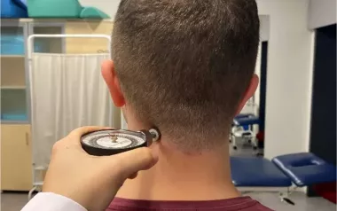
It will be promising to explore specific psychological health impairments from a fascial tissue perspective. A related novel scientific investigation reports about a first exploration in this direction. This study, published by several German scientists, revealed an increased myofascial stiffness of the posterior neck and thoracic erector spinae area in patients with major depressive disorder, compared to healthy controls.40 Interestingly, these tissues also had a less elastic recoil capacity, also described as higher viscoelastic creep (Image 6). 
This surprising finding—at least from a scientific perspective—stimulates new research into several novel questions: To what part is the increased stiffness influenced by muscular fibers and/or to fascial morphology? Is the increased creep capacity related to an increased inflammatory milieu? Or to the “character armor” concept described by Wilhelm Reich?41 How much does the common head-forward posture reflect or influence these tissue changes? Or the less bouncy quality of their gait? And of course: Could a skilled myofascial tissue manipulation serve as a valuable add-on in a related therapeutic program? 
In fact, the same study explored the latter question via an administered self-myofascial foam rolling application to the posterior neck and middle thoracic regions. When compared with a placebo foam rolling treatment, this therapeutic intervention was shown to elicit a reduced negative memory bias and more positive affect in these patients. Do not mistake this effect for a temporary mood swing. Instead, the related test used by our research group measures their perceptual filter, which indicates a much more fundamental cortical process. When hearing a long list of words, depressive patients mostly remember (or recognize again) the negative ones when hearing them again a few minutes later. Long-term psychotherapy, as well as effective pharmacological treatments, have been positively tested to change this filtering action in depressive patients. That a mechanical myofascial self-stimulation now expresses a similar effect indicates a profound reorientation for this field. To give an example: Imagine you take a depressive friend for a five-hour beautiful nature walk, which you have prepared for weeks. At the end, he tells you there was one tiny negative experience, like a very small piece of paper thrown away on the path by a child or a tiny sting by an ant, that spoiled the whole walk for him. How would this influence your motivation to proceed? And could it be that a leisurely walk for a few minutes near your home would be much better for him, if he preceded that with a suitable form of myofascial stimulation?
What else is known about the effect of myofascial stimulation on psychological factors or the ANS? Since Ruffini endings are particularly sensitive to a slow mechanical stimulation with vectorial variations (see the “local listening” concept described before), myofascial release treatment methods and deep-tissue manipulation modalities, such as the Rolfing method of structural integration, may therefore involve a focused stimulation of these fascial mechanoreceptors. It is therefore interesting to note that the stimulation of Ruffini receptors has been shown to elicit a long-lasting effect on the ANS in terms of an inhibited sympathetic activation and augmented parasympathetic activation.42 In congruence with this general physiological expectation, clinical measurement of heart rate variability in human patients before and after a myofascial treatment application (here based on the Rolfing method) revealed a significant parasympathetic shift in the treated patients.43 
No less exciting is a novel publication from Brazil, in which a controlled local inflammation was induced in the lumbar fascia of mice. A gentle myofascial massage was subsequently applied to some of the animals, while the others received no such treatment. The study revealed that the myofascial stimulation elicited a clear anti-inflammatory effect on the local tissue, based on the altered expression of the cytokines IL-4 and TGF-ß1.44 If applicable to human patients, this suggests that gentle myofascial massage stimulation could influence the dynamic interplay between fascial biochemistry, nociception, and immune system regulation. Not a complete surprise: The findings of this new Brazilian study will be the focus of a major keynote presentation and related discussions at the upcoming 6th International Fascia Research Congress in Montreal in September 12–14, 2022. 
Ultimately, we can see that the body-wide fascial system is a rich sensory network—not only because of its newly determined quantity of 250 million nerve endings, but also because of the richness of other important health aspects that are interacting with this sensory network, ranging from the ANS to tissue stiffness regulation to immune system functioning and psycho-emotional factors. 

 Robert Schleip, PhD, is director of the Fascia Research Project (Ulm University and Technical University of Munich in Germany), vice president of the Fascia Research Society, and research director of the European Rolfing Association. His clinical background includes more than 25 years as a teacher and practitioner of the Rolfing method of structural integration and also of the Feldenkrais method of somatic education. He had authored numerous publications in the fascia research field, and his laboratory research on active fascial contractility was given the Vladimir Janda Award for Musculoskeletal Medicine. Learn more at www.somatics.de and www.fasciaresearch.de.