Scoliosis

Clinical Orthopedic Manual Therapy Treatment

By Joseph E. Muscolino, DC
[Feature]

Viewed from posterior to anterior, the human spine should be straight. Any frontal plane curve seen from this view is described as a scoliosis. Therefore, by definition, scoliosis is a lateral flexion deformity of the spine. If one scoliotic curve is present, it is known as a C-curve. If two curves are present, it is known as an S-curve. And if three curves are present, it is known as a double S-curve (Images 1A–1C). When naming a scoliotic curve, it is named for the side of convexity. For example, the curve seen in Image 1A is a right lumbar C-scoliosis; Image 1B demonstrates a right lumbar, left thoracic S-scoliosis; and Image 1C shows a right lumbar, left thoracic, and right cervicothoracic double S-scoliosis. Further, when a scoliotic curve occurs in the frontal plane, there is also a concomitant transverse plane rotation to the curve.
In the lumbar spine, lateral flexion to one side is accompanied by rotation to the opposite side (likely due to the locked-short transversospinalis musculature on the side of concavity). The right C-curve seen in Image 1A comprises lateral flexion to the left, but rotation to the right (rotation is always named for where the anterior aspect of the vertebral body orients). One complication of this is that the spinous processes rotate into the concavity so that the degree of the scoliotic curve is usually greater than what is appreciated by visualizing or palpating the spinous processes. To fully see the degree of a lumbar scoliotic curve, an X-ray is needed.

Effect of Scoliosis

As with any postural distortion pattern, a scoliotic curve can present as mild, moderate, or severe. The degree that this impacts the client can vary tremendously. In and of itself, a scoliotic curve does not necessarily cause pain or dysfunction. There is often a tremendous lag between objective structural distortion and subjective pain and dysfunction. However, a scoliotic curve does lead to a number of compression and tension forces that will likely lead to dysfunction, if not pain, in the long run. For example, the musculature in the concavity will adaptively shorten and become locked-short. The musculature on the convex side will be lengthened and become locked-long (Image 2). Tightness of musculature often leads to pain due to the tension forces within the musculature itself, as well as on its attachments. Tight musculature will also usually resist lengthening and therefore limit motion of the joints that are crossed. Further, the facet joints and the vertebral bodies on the concave side will become compressed. The same structures on the convex side will be under tension stress. This leads to increased physical stresses on the joints that can cause pain and dysfunction, and perhaps lead to greater osteoarthritic degeneration in time.

Clinical Orthopedic Manual Therapy Treatment

For all these reasons, clinical orthopedic manual therapy treatment can play an important role in the treatment plan for clients who have scoliosis. Treatment should be aimed at both loosening the tight musculature involved and mobilizing the joint dysfunction hypomobilities that occur. Tight musculature can be treated with heat, soft-tissue manipulation (massage), and stretching. Joint hypomobility dysfunction can be treated with heat and joint mobilization.

Soft-Tissue Manipulation

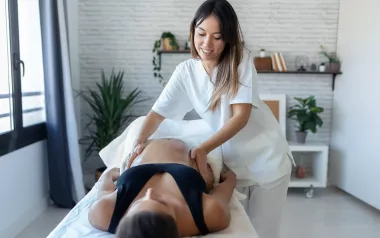
Soft-tissue massage should be oriented at any and all tight myofascial tissue. But usually, greater focus should be placed on the locked-short musculature on the side of the concavity. The primary focus should be the paraspinal musculature—the erector spinae and transversospinalis groups—but the transversospinalis musculature (rotatores, multifidus, semispinalis) in the laminar groove of the spine should be especially focused on. The quadratus lumborum should also be a primary target of massage (Image 3). As with all manual therapy, if the massage is done after heat is applied to soften and warm up the tissue, the work will usually be more effective.
I don’t believe there is any one magical stroke for working on tight musculature. Because locked-short and locked-long muscles tend to be globally tight, deep stripping strokes performed along the length of the musculature can be very effective. Cross-fiber work tends to be better at breaking up patterns of adhesions that would likely develop as the condition becomes more chronic. I like to think of the manual therapist’s job as being a detective, searching for the tight/taut tissues. For this reason, I am a fan of circular strokes because they allow us to approach the tissue from all angles, increasing the likelihood that we will find the tightest areas.
When performing massage, I often think of something that Sandy Fritz, massage therapy educator and author, once said to me: “No massage stroke should ever end the way it was intended when it was begun.” I don’t know if this statement was original to Sandy, but it has stayed with me for many years. This means that instead of performing cookbook strokes, we should amend and adjust them based on the client’s tissues. When receiving massage from a new therapist, this is the first thing I notice. Is the therapist feeling my tissues? Is the therapist adjusting their work to the state of my tissues? Is the therapist adjusting their work to the response of my tissues to the stroke they are performing? The ability to adjust the work to the tissues of the client is the essence of competent clinical orthopedic manual therapy.

Stretching

After heat and/or massage have been performed, we should stretch the client’s tissues. When it comes to stretching a scoliotic spine, it is extremely important that the stretch force is applied to the scoliotic curve itself. The problem is that the scoliotic curve will resist lengthening, and the rest of the spine will tend to compensate and move instead. This is especially true if the client has an S-scoliotic curve (Image 4A). For example, if the client has a lumbar scoliotic curve of left lateral flexion (termed a right lumbar scoliotic curve based on the convexity being on the right side) and we stretch the client into right lateral flexion, if the force is not specifically applied to the lumbar region, the thoracic region will likely move instead. And if the client has a thoracic scoliotic curve in the opposite direction—in other words, a curve of right lateral flexion (termed left scoliotic curve for the convexity on the left)—it is even more likely that the thoracic spine will absorb the stretch and move into right lateral flexion, preventing the stretch from occurring in the lumbar region (Image 4B). In fact, not only will the stretch not be effective for helping the lumbar scoliotic curve, it will actually worsen the thoracic scoliotic curve. The reverse of this is also true. If we stretch the client’s spine into left lateral flexion with the intent of lengthening out the thoracic scoliotic curve, if not specifically applied to the thoracic region, the lumbar region will move instead, both allowing the thoracic curve to avoid being stretched, and worsening the lumbar scoliotic curve (Image 4C).
For this reason, stretching applied to the client’s spine for scoliosis must be done with attention, specificity, and care. If we are giving the client self-care stretches to do at home, we need to instruct them and work with them to stretch the needed region of the spine. Looking at the same examples as in Images 4A–4C, Images 5A–5C demonstrate effective application of lateral flexion stretching in each direction.

Treating the Underlying Cause of Scoliosis

Direct treatment of the scoliosis itself is extremely valuable. However, there may be an underlying cause of scoliosis that, if not addressed, will lead to a perpetuation of the scoliotic curve regardless of the manual therapy treatment performed for the tissues at the scoliotic curve. For example, if the client has a dropped arch due to overpronation of the subtalar joint, the length of the lower extremity on that side will decrease, leading to a dropped pelvis on that side. If the spine were to remain straight, it would resemble the Leaning Tower of Pisa (Image A), which would result in the eyes and inner ears being unlevel, making proprioception difficult. To prevent this, the spine would develop a compensatory scoliotic curve to bring the head to a level posture (Image B).
In a case like this, direct treatment of the scoliosis itself is treating the symptom, not the cause. This is not to say that treatment of the locked myofascial tissues and joint dysfunction of the scoliosis is not needed—it very much is. But it will never truly resolve the issue if the underlying cause, in this case the dropped arch, is not also resolved. Similarly, any condition that causes the pelvis to drop on one side, such as genu valgum of the knee or asymmetrical tightness of frontal plane abduction/adduction musculature of the hip joint, would likely result in a compensatory scoliosis.
Of course, not every scoliosis is caused by dysfunction in the kinematic chain of the lower extremity. Some scolioses are described as “idiopathic” because the cause of the condition is unknown (“idio” meaning unknown, and “path” referring to the condition). But because there often is an underlying structural cause of the scoliosis, it is important to assess for its possible presence, and if found, treat it.

Stretching

Stretching is a simple concept: it is done to make soft tissues longer. Stretching is essentially a mechanical process, although neural inhibition stretching techniques that make use of the reciprocal inhibition neurological reflex and/or Golgi tendon organ reflex can also be done. These neural techniques have many names and many abbreviations: agonist contract (AC), contract relax (CR), proprioceptive neuromuscular facilitation (PNF), PIR, AIS, MET, etc. Although there is a great deal of controversy lately about the effectiveness of stretching, there is a fundamental characteristic of soft tissue known as creep. That is, a sustained force placed on a soft tissue will cause the soft tissue to deform to that force. In this case, the deforming—in other words, changing form—is to have a short soft tissue become longer, or at least more flexible so that it can lengthen.

Using Bolsters to Stretch and Treat

When stretching the client in our office, we can take advantage of bolsters to focus the stretch to the scoliotic curve. For example, if the client has a right lumbar scoliosis, we would place them in side-lying position on their right side (Image 6A). We would then place the bolster ideally under the apex of the lumbar scoliotic curve (Image 6B). If they have a left thoracic scoliosis, we would place them in side-lying position on their left side with the bolster under the apex of the thoracic scoliotic curve (Image 6C). In either case, we place the client side-lying with the convexity of the scoliosis oriented down against the table. The force of the bolster pushing up against the spine would stretch and reduce the scoliotic curve. We have the choice of using smaller or larger bolsters and softer or firmer bolsters depending on the force of the stretch we want, and what the client’s body can comfortably allow.

Add in Stretch from the Extremities

The effectiveness of this stretch can be increased if we add in a tension stretch force from the extremities (Images 7A and 7B). Image 7A shows the client’s thigh dropped down off the backside of the table into adduction and the pelvis brought down into depression. This would increase the stretch to the lumbar scoliotic curve. In fact, the effectiveness of the bolster plus dropping the lower extremity off the table, is such that the client’s lumbar scoliotic curve is actually a bit overcorrected, or curved in the opposite direction in the frontal plane. This provides an even stronger stretch so that when the corrections are removed, the improvement to the scoliotic curve should be even greater. Image 7B shows the client’s upper extremity and shoulder girdle stretched down to increase the stretch to the thoracic scoliotic curve.

Lateral Flexion-Break Table

If you have a table with what is known as a “flexion-break” feature, it can be used as a “lateral flexion-break” to stretch the scoliotic curves in a similar fashion to using bolsters. The break in the table would be placed at the apex of the scoliotic curve and the table would be brought into lateral flexion to the desired degree (Image 8). This is easier logistically for the therapist than working with bolsters because the therapist can change the degree of stretch with the touch of the foot pedal, whereas changing the stretch force with bolsters would require the client to get up and down on the table each time the stretch force is changed by changing the size of the bolster. Gently increasing the force of the stretch over time to allow the client’s tissues to accommodate and accept the stretch is advised. Begin the client with a gentle degree of lateral flexion, maintaining them in this position for perhaps 10–30 seconds. Once the client is comfortable in this position, incrementally increase the degree of lateral flexion of the table, each time maintaining the client in that position for enough time to allow their tissues to accept the stretch force. Of course, a bolster could be added to this technique and stretching the lower or upper extremity could also be added to increase the assertiveness of the stretch.

Joint Mobilization

As effective as stretching can be to reduce the degree of a scoliotic curve, it may not be sufficiently effective at stretching some of the tight/taut myofascial tissues of the client’s spine because the stretch force is spread out over the entire region of the scoliotic curve. If a more specific stretch force is needed, perhaps to stretch the tissues at a specific segmental joint level of the spine, Grade IV joint mobilization techniques can be used instead (see “Grade IV Joint Mobilization,” below). Grade IV joint mobilization of the spine, performed with slow oscillations, is a form of joint stretching that is much more specifically applied so that it better targets the intrinsic fascial tissues of the facet joints of the spine at a specific level. For this reason, joint mobilization might be better termed arthrofascial stretching. Joint mobilization of the spine also targets the intrinsic musculature of the spine such as the rotatores, intertransversarii, and interspinales.
There are many different joint mobilization techniques that may be directed toward mobilizing the client’s scoliosis. Image 9 demonstrates a side-lying technique. The client is placed in side-lying position on her left side so that the convexity of the right lumbar scoliotic curve is oriented up toward the ceiling. The therapist uses his pisiform to contact the client’s lumbar spinous process/lamina at the desired joint level, trapping the bone against his contact. The therapist then gently sinks down with his body weight. Once tissue tension is reached, the therapist gently increases the force downward toward the table to stretch/mobilize the intrinsic fascial tissue of the spine at that level. Repetitions at that joint level can be performed as desired. This can then be repeated at other levels of the spine.
Another method of joint mobilization of the spine can be performed with the client seated (Images 10A–10C). The client is seated with the convexity of the scoliotic curve oriented toward the therapist. The therapist contacts the spinous process at the desired joint level with either the thumb or pisiform, whichever is comfortable for therapist and client (Image 10A). The therapist now gently but firmly grasps the client’s shoulders/trunk with his other hand. With the contact against the spinous process, the therapist brings the client’s spine into lateral flexion toward him until tissue tension is reached (Image 10B). A gentle mobilization force is added with the left hand or with the right thumb contact (Image 10C). This technique can be repeated as needed at that level and then performed at the other levels of the scoliotic curve.

Grade IV Joint Mobilization

It is interesting to note that “regular” stretching of a joint that applies the stretch force to the end of the client’s passive range of motion can technically be described as Grade III joint mobilization, because stretching is a form of joint mobilization. Grade IV joint mobilization is different because it is much more specifically applied and is usually thought of as increasing the nonaxial glide motion of a joint.
To perform Grade IV joint mobilization, the therapist must locate the specific level to be mobilized and apply the stretch force to that level. As with any stretching, joint mobilization is performed once the tissue is brought to tension, meaning to the tissue-tension mechanical barrier at end range of motion. All slack in the superficial tissues (skin, subcutaneous fascia, and any musculature between the skin and the bone being contacted) must be first removed so that the bone is securely contacted under the therapist’s hand. This aspect of Grade IV joint mobilization is often a bit challenging for massage therapists because they are accustomed to directing their attention to the soft tissues between the skin and bone, not pushing through those soft tissues to “trap” the bone against their contact. Once this contact is achieved, the mobilization force is gently applied with oscillation motions, usually a millimeter or two and held for only a fraction of a second. As with massage or regular stretching, there is no hard-and-fast rule for how many oscillation repetitions to perform. One good guideline is to perform the mobilization technique for sets of 3–5 mobilizations or perhaps sets of 10–15 seconds of repetitions.

Ethics and Legality

Note: Please make sure you are legally and ethically allowed within the scope of your license/certification to practice any manual therapy technique, including Grade IV joint mobilization, before employing it in your practice. Further, no kinesthetic technique should be added to your practice unless you have had in-person training with a professional continuing education instructor skilled in that technique. It is important to emphasize that Grade IV joint mobilization is employed with slow oscillations, never a fast thrust. A fast thrust applied to joint stretching defines a Grade V joint mobilization, which is only legally and ethically allowed for certain manual therapy professions, such as chiropractic and osteopathic physicians and physical therapists.

Precautions and Contraindications

Every technique has precautions and contraindications. Joint mobilization is contraindicated if there is hypermobile/unstable tissue at the region being mobilized (including, but not limited to, a herniated disc or osteoporosis).

Joseph E. Muscolino, DC, has been a manual and movement therapy educator for more than 30 years. He is the author of multiple textbooks, including The Muscular System Manual: The Skeletal Muscles of the Human Body (Elsevier, 2017); The Muscle and Bone Palpation Manual with Trigger Points, Referral Patterns, and Stretching (Elsevier, 2016); and Kinesiology: The Skeletal System and Muscle Function (Elsevier, 2017). He is also the author of 12 DVDs on manual and movement therapy and teaches continuing education workshops around the world, including a certification in Clinical Orthopedic Manual Therapy (COMT), and has created Digital COMT, a video streaming subscription service for manual and movement therapists, with new content added each and every week. Visit www.learnmuscles.com for more information or reach him directly at joseph.e.muscolino@gmail.com.